 Indietro
Anatomy & Physiology Study Guide: The Respiratory System
Guida di studio - Note intelligenti
 Appunti personalizzati basati sui tuoi materiali, ampliati con definizioni chiave, esempi e contesto.
Respiratory System Overview
Introduction
The respiratory system is responsible for gas exchange, supplying oxygen to the blood and removing carbon dioxide. It is divided into upper and lower respiratory tracts, each with distinct anatomical structures and functions. 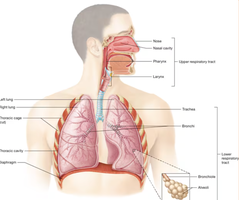
Upper Respiratory Tract
Nasal Cavity
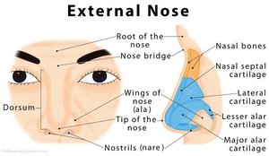
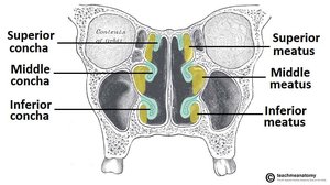
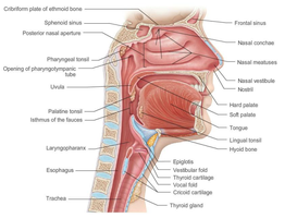
The nasal cavity is the entry point for air and plays a key role in filtering, warming, and humidifying inhaled air. - Nares: The nostrils, which are the external openings. - Vestibule: The entrance to the nasal cavity. - Nasal septum: The wall dividing the nostrils. - Nasal conchae: Curved bony structures (superior, middle, inferior) that increase surface area. - Meatuses: Passages beneath each concha. 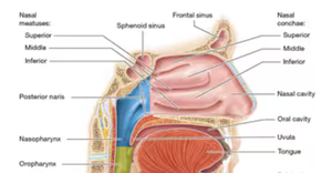
Paranasal Sinuses & Pharynx
Paranasal sinuses are air-filled spaces that lighten the skull and help warm and moisten air. The pharynx is a muscular tube connecting the nasal cavity to the larynx and esophagus. - Sinuses: Frontal, sphenoid, maxillary, and ethmoid. - Pharynx: Divided into nasopharynx, oropharynx, and laryngopharynx. 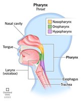
Larynx
The larynx, or voice box, is involved in sound production and protects the lower airways. - Laryngeal prominence: Commonly known as the "Adam's apple." - Epiglottis: Prevents food from entering the trachea. - Vocal folds: Produce sound. 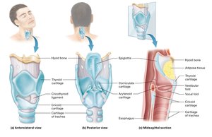
Lower Respiratory Tract
Trachea
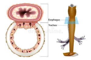
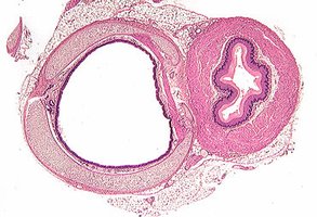
The trachea is a tube supported by C-shaped cartilage rings, conducting air to the bronchi. - Carina: The point where the trachea splits into right and left primary bronchi. - Esophagus: Located posterior to the trachea, mainly muscular. 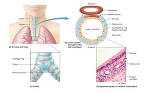
Trachea Histology
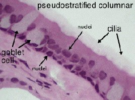
The trachea consists of several layers, each with specialized functions. - Mucosa: Ciliated pseudostratified columnar epithelium with goblet cells producing mucus. - Submucosa: Connective tissue with seromucous glands. - Hyaline cartilage: Provides flexible support. - Adventitia: Anchors trachea to surrounding structures. 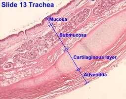
Bronchi and Bronchial Tree
The bronchial tree branches from the trachea into progressively smaller airways. - Primary bronchi: Right and left main bronchi. - Secondary (lobar) bronchi: Supply each lung lobe. - Tertiary (segmental) bronchi: Supply bronchopulmonary segments. 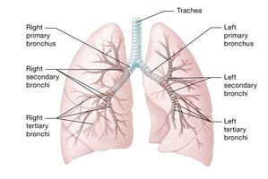
Lungs
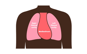
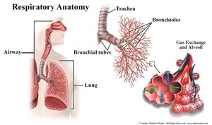
The lungs are paired organs with distinct anatomical features. - Right lung: Superior, middle, and inferior lobes; horizontal and oblique fissures. - Left lung: Superior and inferior lobes; oblique fissure; cardiac notch. - Apex: Top of the lung. - Base: Bottom of the lung. - Mediastinum: Central region separating the lungs. 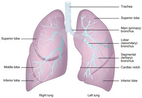
Hilum of the Lung
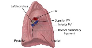
The hilum is the area where bronchi, pulmonary arteries and veins, and nerves enter and exit the lung. 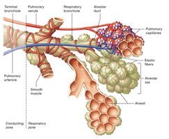
Alveoli of the Lungs
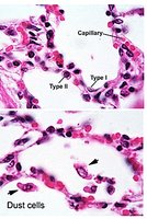
Alveoli are tiny air sacs where gas exchange occurs. - Type 1 alveolar cells: Squamous cells for gas exchange. - Type 2 alveolar cells: Cuboidal cells producing surfactant. - Alveolar macrophages: "Dust cells" that remove debris. 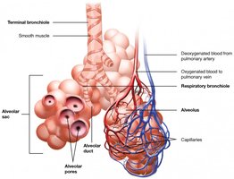
Muscles of Inspiration and Expiration
Mechanics of Breathing
Breathing involves changes in thoracic volume and pressure, governed by Boyle’s Law. - Inspiration: Increase in volume, decrease in pressure, air flows in. - Expiration: Decrease in volume, increase in pressure, air flows out. Boyle’s Law: Key Muscles: - Inspiration: Diaphragm, external intercostals - Expiration: Internal intercostals, abdominal muscles
Spirometry and Pulmonary Function
Spirometry Activity
Spirometry measures lung volumes and capacities to assess pulmonary function. Directly Measured Volumes: - Tidal Volume (TV): Air moved in/out during normal breathing. - Inspiratory Reserve Volume (IRV): Extra air inhaled after normal inspiration. - Expiratory Reserve Volume (ERV): Extra air exhaled after normal expiration. - Vital Capacity (VC): Maximum air exhaled after maximum inhalation. - Inspiratory Capacity (IC): TV + IRV Formulas: Obstructive Lung Disease: Difficulty exhaling (e.g., asthma, COPD). Restrictive Lung Disease: Difficulty inhaling (e.g., fibrosis).
Forced Vital Capacity and FEV1
- Forced Vital Capacity (FVC): Total air forcefully expelled during exhalation. - Forced Expiratory Volume in 1 second (FEV1): Air expelled in the first second of forced exhalation.
Histology Review and Lab Quiz Preparation
Key Histological Features
- Trachea: Goblet cells and cilia in pseudostratified columnar epithelium. - Lung: Type 1 vs. type 2 alveolar cells, dust cells. Lab Preparation: - Identification of structures on models. - Definitions of spirometry terms and tissue types.
Summary Table: Respiratory System Structures
Structure | Main Function | Key Features |
|---|---|---|
Nasal Cavity | Filters, warms, humidifies air | Conchae, meatuses, vestibule |
Pharynx | Passage for air and food | Nasopharynx, oropharynx, laryngopharynx |
Larynx | Sound production, airway protection | Vocal folds, epiglottis, cartilage |
Trachea | Conducts air to bronchi | C-shaped cartilage, mucosa, cilia |
Bronchi | Air distribution | Primary, secondary, tertiary branches |
Lungs | Gas exchange | Lobes, fissures, hilum, alveoli |
Alveoli | Gas exchange | Type 1 & 2 cells, macrophages |
Additional info:
- The mediastinum is the central compartment of the thoracic cavity, containing the heart, great vessels, and other structures. - Surfactant produced by type 2 alveolar cells reduces surface tension, preventing alveolar collapse. - Spirometry is essential for diagnosing and monitoring respiratory diseases. ----------------------------------------
