 Indietro
Autonomic Regulation of the Eye, Auditory System, Muscle Physiology, and Cardiac Function
Guida di studio - Note intelligenti
 Appunti personalizzati basati sui tuoi materiali, ampliati con definizioni chiave, esempi e contesto.
Autonomic Regulation of the Eye
Pupillary Constriction and Dilation
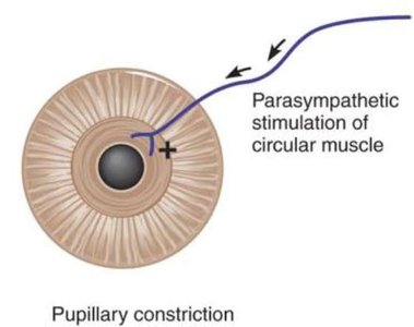
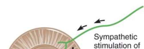
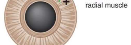
The diameter of the pupil is controlled by the autonomic nervous system, which regulates the activity of two types of smooth muscle in the iris: the circular muscle and the radial muscle. This control is essential for adjusting the amount of light entering the eye.
Parasympathetic stimulation activates the circular muscle, causing pupillary constriction (miosis).
Sympathetic stimulation activates the radial muscle, causing pupillary dilation (mydriasis).
This antagonistic control allows the pupil to respond to changes in ambient light and emotional states.
Example: Pupillary constriction occurs in bright light, while dilation occurs in dim light or during fight-or-flight responses.

The Auditory System
Anatomy and Function of the Ear
The ear is responsible for detecting sound waves and converting them into neural signals. The process involves the transmission of sound through the external, middle, and inner ear, culminating in the activation of hair cells in the cochlea.
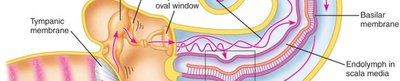
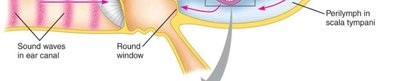
Sound waves enter the ear canal and vibrate the tympanic membrane.
Vibrations are transmitted via the ossicles (malleus, incus, stapes) to the oval window.
Movement of the oval window creates waves in the perilymph and endolymph of the cochlea.
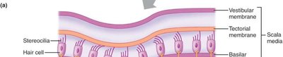
These waves cause displacement of the basilar membrane, stimulating hair cells.
Hair cells transduce mechanical energy into electrical signals, which are sent to the brain via the cochlear nerve.
Example: The basilar membrane's movement is frequency-dependent, allowing pitch discrimination.

Muscle Physiology
Muscle Cell Structure and Terminology
Muscle tissue is composed of specialized cells and structures that enable contraction. Understanding the terminology is essential for studying muscle function.
Muscle cell: Also called a muscle fiber.
Sarcolemma: Plasma membrane of a muscle fiber.
Triad: Structure formed by a T tubule and two lateral sacs of the sarcoplasmic reticulum.
End-plate potential (EPP): Graded potential that excites skeletal muscle cells.
Fascicles: Bundles of muscle fibers divided by perimysium.
Lateral sac: Enlargement of the sarcoplasmic reticulum where calcium is stored.
Endomysium: Thin sheath of connective tissue surrounding individual muscle fibers.
Sarcoplasmic reticulum (SR): Modified smooth ER that stores calcium.
Epimysium: Connective tissue surrounding the whole muscle.
Motor unit: Somatic motor neuron and the muscle fibers it innervates.
Myofibril: Banded, rodlike elements containing contractile machinery.
T tubules: Deep indentations in sarcolemma transmitting action potentials.
Muscle Contraction Mechanisms
Muscle contraction is driven by the interaction of thick and thin filaments within the sarcomere, regulated by various proteins and structures.
Sliding filament model: Describes how actin and myosin filaments slide past each other to produce contraction.
Sarcomere: The smallest functional unit of muscle fibers, defined by Z lines.
Crossbridge cycling: The repeated attachment and detachment of myosin heads to actin filaments.
Actin: Contractile protein making up the thin filament.
Myosin: Contractile protein making up the thick filament.
Tropomyosin: Regulatory protein that blocks myosin-binding sites on actin.
Troponin: Regulatory protein that binds calcium and moves tropomyosin off myosin-binding sites.
DHP receptors: Voltage sensors in T tubule membrane.
Ryanodine receptors: Calcium channels in SR membrane.
Example: Calcium release from SR triggers contraction by enabling crossbridge formation.
Muscle Types Comparison
Muscle tissue is classified into skeletal, smooth (single-unit and multi-unit), and cardiac types, each with distinct properties.
Property | Skeletal | Smooth (single unit) | Smooth (multi-unit) | Cardiac |
|---|---|---|---|---|
Striations (sarcomeres) | Yes | No | No | Yes |
Actin and myosin | Present | Present | Present | Present |
Level of control | Voluntary | Involuntary | Involuntary | Involuntary |
Neural input | Somatic | Autonomic | Autonomic | Autonomic |
Neuroeffector junction | Motor end plate | Diffuse | Diffuse | Intercalated discs |
Hormonal control | No | Yes | Yes | No |
Source of calcium | Sarcoplasmic reticulum | SR and extracellular | SR and extracellular | SR and extracellular |
Regulatory protein that binds with calcium | Troponin | Calmodulin | Calmodulin | Troponin |
Gap junctions | No | Yes | No | Yes |
Pacemaker activity | No | Yes | No | Yes |
Myosin ATPase activity | High | Low | Low | Intermediate |
























Cardiac Function
Heart Anatomy and Blood Flow
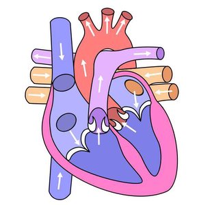
The heart is a muscular organ responsible for pumping blood throughout the body. It consists of four chambers: two atria and two ventricles. Blood flow is regulated by valves and follows a specific pathway through the heart and circulatory system.
Right atrium receives deoxygenated blood from the body.
Right ventricle pumps blood to the lungs for oxygenation.
Left atrium receives oxygenated blood from the lungs.
Left ventricle pumps oxygenated blood to the body.
Valves prevent backflow and ensure unidirectional flow.
Example: The pulmonary and aortic valves regulate blood flow from the ventricles to the arteries.
Cardiac Action Potential
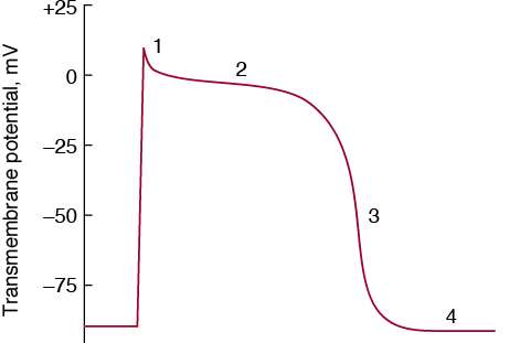
Cardiac muscle cells generate action potentials that coordinate contraction. The action potential has distinct phases, each associated with specific ion movements.
Phase 0: Rapid depolarization due to Na+ influx.
Phase 1: Initial repolarization.
Phase 2: Plateau phase due to Ca2+ influx balancing K+ efflux.
Phase 3: Repolarization due to K+ efflux.
Phase 4: Resting membrane potential.
Example: The plateau phase is unique to cardiac muscle and prevents tetanus.
Additional info: The notes above expand on the original content by providing definitions, examples, and context for each topic, ensuring completeness and academic quality for exam preparation.
