 Indietro
Blood: Structure, Function, and Disorders – Study Notes for Anatomy & Physiology
Guida di studio - Note intelligenti
 Appunti personalizzati basati sui tuoi materiali, ampliati con definizioni chiave, esempi e contesto.
Blood: Structure, Function, and Disorders
Functions of Blood
Blood is a vital connective tissue responsible for multiple essential functions in the human body.
Transport: Carries oxygen, nutrients, wastes, and hormones throughout the body.
Regulation: Maintains body temperature, pH balance, and fluid volume.
Protection: Prevents blood loss (hemostasis) and fights infections via immune cells.
Composition and Physical Characteristics of Blood
Blood consists of plasma (the fluid matrix) and formed elements (cells and cell fragments).
Plasma: Nonliving fluid matrix, makes up ~55% of blood volume.
Formed Elements: Living cells suspended in plasma, including erythrocytes (RBCs), leukocytes (WBCs), and platelets.
Hematocrit: Percentage of blood volume occupied by RBCs; normal values are 47% ± 5% for males and 42% ± 5% for females.
Physical Properties: Sticky, opaque fluid with metallic taste; color varies with oxygen content (scarlet red for high O2, dark red for low O2); pH 7.35–7.45; makes up ~8% of body weight.
Erythrocytes (Red Blood Cells)
Erythrocytes are specialized for oxygen transport and exhibit unique structural features.
Structure: Small (7.5 μm), biconcave, anucleate, lacking most organelles; filled with hemoglobin (Hb).
Function: Efficient gas transport due to large surface area, high Hb content, and anaerobic metabolism (do not consume O2).
Key Functions:
Carry oxygen from lungs to tissues.
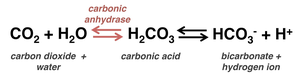
Release carbonic anhydrase into plasma to facilitate CO2 transport to lungs.
Act as acid-base buffers to regulate blood pH.
Hemoglobin and Oxygen Transport
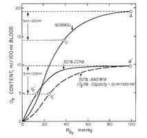
Hemoglobin is the protein responsible for oxygen binding and transport in erythrocytes.
Each Hb molecule: Can bind four O2 molecules.
O2 loading: In lungs, forms oxyhemoglobin (ruby red).
O2 unloading: In tissues, forms deoxyhemoglobin (dark red).
CO2 transport: 20% of CO2 binds to Hb as carbaminohemoglobin.
Erythropoiesis: Formation of Red Blood Cells
Erythropoiesis is the process of RBC production, regulated by hormones and dietary factors.
Stages: Hematopoietic stem cell → myeloid stem cell → proerythroblast → erythroblast stages → reticulocyte → mature erythrocyte.
Regulation: Erythropoietin (EPO) stimulates RBC production; testosterone increases EPO production.
Dietary Requirements: Amino acids, lipids, carbohydrates, iron (bound to ferritin, hemosiderin, transferrin), vitamin B12, and folic acid.
Erythrocyte Disorders
Disorders of erythrocytes include anemia (low O2-carrying capacity) and polycythemia (excess RBCs).
Anemia: Classified by cause:
Blood loss: Hemorrhagic anemia (acute or chronic).
Not enough RBCs produced: Iron-deficiency, pernicious, renal, and aplastic anemia.
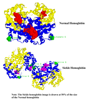
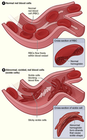
Too many RBCs destroyed: Hemolytic anemias (e.g., thalassemias, sickle-cell anemia).
Polycythemia: Excess RBCs increase blood viscosity; causes include polycythemia vera (bone marrow cancer), secondary polycythemia (high altitude), and blood doping.
Leukocytes (White Blood Cells)
Leukocytes are immune cells that defend against disease and are classified as granulocytes or agranulocytes.
Granulocytes: Neutrophils, eosinophils, basophils (contain visible granules).
Agranulocytes: Lymphocytes, monocytes (lack visible granules).
Leukocytosis: Elevated WBC count (>11,000/μl), normal response to infection.
Neutrophils
Most numerous WBC; first responders to infection; highly phagocytic.
Kill microbes via respiratory burst (producing bleach or hydrogen peroxide).
Defensin granules pierce membranes of ingested microbes.
Eosinophils
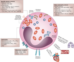
Primarily combat parasitic infections and mediate allergic responses.
Secrete chemokines, cytokines, and growth factors for inflammation and parasite destruction.
Basophils
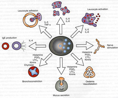
Responsible for allergic and antigen responses; release histamine and heparin.
Histamine increases blood vessel permeability.
Heparin inhibits clotting and promotes WBC movement.
Attract eosinophils and neutrophils to infection sites.
Lymphocytes
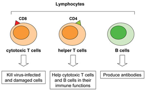
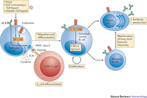
Key players in adaptive immunity; include B cells and T cells.
B cells: Produce antibodies, activate complement system.
T cells:
CD4+ helper T cells: Coordinate immune response via cytokines.
CD8+ cytotoxic T cells: Kill virus-infected and tumor cells.
Monocytes
Circulate in blood, then mature into macrophages or dendritic cells in tissues.
Phagocytose microorganisms and debris.
Present antigens to activate T lymphocytes.
Produce cytokines (e.g., TNF, IL-1, IL-12).
Can kill infected host cells via antibody-dependent cell-mediated cytotoxicity.
Leukocyte Disorders
Disorders include leukemias (overproduction of abnormal WBCs), leukopenia (low WBC count), and infectious mononucleosis.
Leukemias: Cancerous proliferation of abnormal WBCs; classified as myeloid or lymphocytic, acute or chronic.
Leukopenia: Abnormally low WBC count, often drug-induced.
Infectious mononucleosis: Viral disease caused by Epstein-Barr virus; results in enlarged lymphocytes.
Platelets and Hemostasis
Platelets are cell fragments essential for blood clotting and hemostasis.
Formation: Derived from megakaryocytes; regulated by thrombopoietin.
Function: Form temporary platelet plugs to seal vessel breaks; contain chemicals for clotting.
Hemostasis: Three steps: vascular spasm, platelet plug formation, coagulation.
Coagulation Pathways
Coagulation reinforces platelet plug with fibrin threads, transforming blood from liquid to gel.
Uses clotting factors (I–XIII), many requiring vitamin K.
Intrinsic and extrinsic pathways converge to activate prothrombin to thrombin, which converts fibrinogen to fibrin.
Blood Transfusions and Blood Groups
Blood transfusions require careful matching of blood groups to prevent adverse reactions.
ABO Blood Groups: Types A, B, AB, O based on presence of agglutinogens (antigens) on RBCs.
Rh System: Rh-positive (D antigen present) or Rh-negative; anti-Rh antibodies form after exposure.
Transfusion Reactions: Occur if mismatched blood is infused; can cause agglutination, hemolysis, and renal failure.
Universal Donor: Type O; Universal Recipient: Type AB (with caveats).
Diagnostic Blood Tests
Blood tests provide valuable information about health and disease.
Hematocrit: Indicates anemia.
Blood glucose: Screens for diabetes.
Differential WBC count: Helps diagnose infections and blood disorders.
Prothrombin time and platelet counts: Assess hemostasis.
Complete blood count (CBC): Evaluates formed elements, hemoglobin, and hematocrit.
Developmental Aspects of Blood
Blood cell formation changes during development and aging.
Fetal blood cells form in yolk sac, liver, and spleen; red bone marrow becomes primary site by seventh month.
Fetal hemoglobin (Hb F) has higher O2 affinity than adult hemoglobin (Hb A).
Blood diseases of aging include chronic leukemias, anemias, and clotting disorders, often linked to cardiovascular or immune system disorders.
