 Indietro
Bones & Bone Structure: Anatomy and Physiology Study Guide
Guida di studio - Note intelligenti
 Appunti personalizzati basati sui tuoi materiali, ampliati con definizioni chiave, esempi e contesto.
Bones & Bone Structure
Functions of the Skeletal System
The skeletal system is composed of bones, cartilages, ligaments, and other connective tissues. It serves several essential functions in the human body:
Support: Provides a structural framework for the body.
Storage of minerals & lipids: Stores calcium, phosphate, and fat in bone marrow.
Blood cell production (hematopoiesis): Occurs in red bone marrow.
Calcium regulation: Maintains blood calcium levels.
Protection: Shields vital organs (e.g., skull protects the brain).
Leverage/movement: Bones act as levers for muscles to produce movement.
Classification of Bones by Shape
Bones are classified according to their shape, which relates to their function and location:
Long Bones: Found in arms, legs, palms, soles, fingers.
Flat Bones: Roof of skull, sternum, ribs, scapulae.
Short Bones: Carpals (wrist), tarsals (ankle).
Irregular Bones: Vertebrae, pelvis, some skull bones.
Sesamoid Bones: Patella, small bones near joints.
Sutural Bones: Small, irregular bones found between skull sutures; vary in number, shape, and position.
Bone Structure
Structure of a Long Bone
Long bones have a distinct anatomy that supports their function:
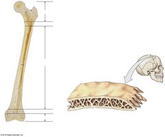
Diaphysis (shaft): The central, elongated portion; contains the medullary (marrow) cavity.
Epiphysis (ends): Proximal and distal ends, mostly spongy bone.
Metaphysis: Region between diaphysis and epiphysis; contains the growth plate (cartilage plate).
Articular Cartilage: Hyaline cartilage covering joint surfaces; reduces friction.
Periosteum and Endosteum
The periosteum and endosteum are connective tissue membranes that cover bone surfaces:
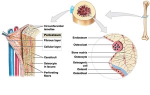
Periosteum: Surrounds and isolates bone; has a fibrous layer (mixes with tendons/ligaments via Sharpey’s fibers) and a cellular layer (growth/repair).
Endosteum: Incomplete layer of osteoprogenitor cells; lines marrow cavity and central canal.
Microscopic Structure: Osteons and Lamellae
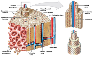
Compact bone is organized into osteons (Haversian systems), which are cylindrical structures:
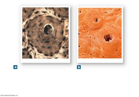
Osteon: Basic unit of compact bone; consists of concentric lamellae surrounding a central canal.
Lamellae: Layers of bone matrix; types include concentric, interstitial, and circumferential lamellae.
Lacunae: Small spaces housing osteocytes.
Canaliculi: Tiny channels connecting lacunae for nutrient/waste exchange.
Types of Bone Tissue
Cortical (Compact) Bone vs. Trabecular (Spongy) Bone
Bones contain two main types of tissue:
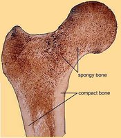
Compact Bone: Dense, forms the external surface; few pores/spaces.
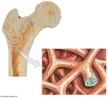
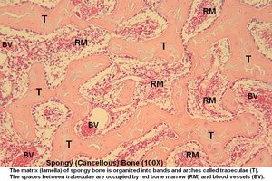
Spongy (Cancellous) Bone: Contains many pores/spaces; organized into trabeculae (bars/plates).
Organization of Compact Bone
Compact bone is highly organized for strength and nutrient delivery:
Osteons: Cylindrical units with concentric lamellae.
Central Canal: Contains blood vessels and nerves.
Volkmann’s (Perforating) Canals: Connect central canals to each other and to the periosteum.
Organization of Spongy Bone
Spongy bone is less organized, with a network of trabeculae:
Trabeculae: Bands and arches of bone matrix.
Red Bone Marrow: Fills spaces between trabeculae; site of hematopoiesis.
Blood Vessels: Supply nutrients to osteocytes.
Bone Marrow
Red vs. Yellow Bone Marrow
Bone marrow is found within the cavities of bones and serves different functions:
Red Bone Marrow: Forms red blood cells; found in proximal epiphysis of humerus/femur, sternum, pelvis, ribs, skull, scapulae, vertebrae.
Yellow Bone Marrow: Stores fat (adipose tissue); energy reserve; found in other bones.
Bone Cells
Types of Bone Cells
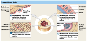
Bone tissue contains several specialized cell types:
Osteoprogenitor Cells: Stem cells that differentiate into osteoblasts; found in endosteum, central canal, periosteum.
Osteoblasts: Bone-forming cells; synthesize organic matrix (osteoid).
Osteocytes: Mature bone cells; maintain bone matrix and calcium homeostasis.
Osteoclasts: Bone-resorbing cells; remove calcium from bone; multinucleated.
Bone Development & Growth
Ossification Processes
Ossification is the process of converting other tissue to bone. There are two main types:
Intramembranous Ossification: Forms bones from embryonic tissue (mesenchyme); occurs in skull, mandible, clavicle.
Endochondral Ossification: Forms bones from hyaline cartilage; most bones develop this way.
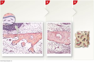
Intramembranous Ossification Steps
Mesenchymal cells cluster and differentiate into osteoblasts.
Osteoblasts lay down osteoid.
Osteoblasts become osteocytes when surrounded by matrix.
Spicules interconnect, trapping blood vessels.
Spongy bone forms, later remodeled to compact bone.
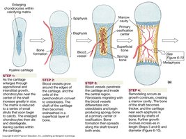
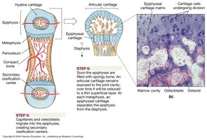
Endochondral Ossification Steps
Bone growth from hyaline cartilage model (~7 weeks development).
Long bones grow through cartilage until puberty.
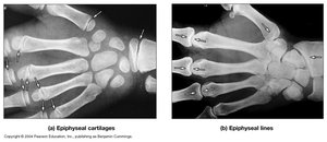
At puberty, cartilage is replaced by bone; epiphyses fuse to diaphysis.
Epiphyseal plate becomes epiphyseal line (ossified).
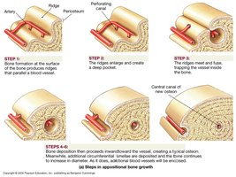
Appositional Bone Growth
Appositional growth increases bone diameter in adults:
Bone formation occurs at the surface, creating new osteons.
Ridges enlarge and fuse, trapping blood vessels.
Bone continues to increase in diameter.
Blood Supply to Bone
Blood Vessels in Bone
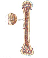
Bones are highly vascularized to support growth and maintenance:
Nutrient arteries: Supply blood throughout diaphysis.
Periosteal artery: Supplies blood to superficial osteons.
Metaphyseal artery: Supplies blood to epiphyseal plate (metaphysis).
Bone Remodeling & Calcium Storage
Bone Remodeling
Bones remodel continuously, with up to 20% replaced per year:
Exercise stimulates osteoblast activity and increases bone density.
Inactivity weakens bone.
Hormones influencing bone growth: growth hormone, calcitriol, sex hormones, thyroxine, calcitonin, parathyroid hormone (PTH).
Calcium Regulation
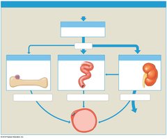
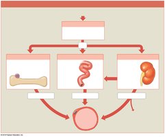
Calcium homeostasis is maintained by bone cells and hormones:
99% of body’s calcium is stored in the skeletal system.
Calcitonin: Decreases blood calcium levels.
PTH: Increases blood calcium levels.
Calcitriol: Works with PTH to absorb calcium from the digestive system; made in kidneys from Vitamin D3.
Fracture Repair
Steps in Fracture Repair
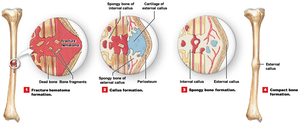
Bone repair depends on adequate blood flow and survival of cells in periosteum/endosteum:
Hematoma forms to control bleeding.
Cells divide to form callus (external: cartilage/bone; internal: spongy bone).
Cartilage replaced by spongy bone.
Extra material is remodeled over months to years.
Fracture Categories
Open (Compound) Fracture: Bone projects through skin.
Closed (Simple) Fracture: Bone remains internal.
Displaced Fracture: Bone fragments out of alignment.
Epiphyseal Fracture: Involves epiphyseal plate; can affect growth.
Common Fracture Patterns
Greenstick Fracture: Incomplete break, common in children.
Spiral Fracture: Caused by twisting forces.
Pott’s Fracture: Ankle fracture.
Colles’ Fracture: Distal radius fracture.
Compression Fracture: Bone is crushed.
Effects of Aging on Bone
Osteopenia and Osteoporosis
Bone density naturally decreases with age:
Osteopenia: Natural decrease in bone density; more common in women (menopause = decreased estrogen).
Osteoporosis: Severe decrease in bone density; affects daily function; risk higher in females, especially postmenopausal women.
Onset can be delayed and progression slowed with calcium, vitamin D, and exercise.
Bones most affected: epiphyses of long bones, vertebrae, mandible, maxilla.
