 Indietro
Bones and Bone Structure: Study Guide for Anatomy & Physiology
Guida di studio - Note intelligenti
 Appunti personalizzati basati sui tuoi materiali, ampliati con definizioni chiave, esempi e contesto.
Bones and Bone Structure
Functions of the Skeletal System
The skeletal system is a complex framework that provides structural support and facilitates movement. It consists of bones, cartilages, ligaments, and other connective tissues.
Support: Provides the body with its shape and supports soft tissues.
Storage of minerals and lipids: Stores calcium, phosphorus, and lipids for energy.
Blood cell production: Houses red bone marrow, which produces blood cells.
Protection: Shields vital organs (e.g., skull protects the brain).
Leverage: Acts as levers for muscles to facilitate movement.
Classification of Bones
Bones are classified by their shape and structure, each serving distinct functional roles.
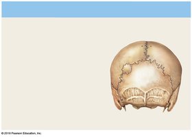
Sutural (Wormian) Bones: Small, flat, irregular bones found between skull bones. Borders resemble jigsaw puzzle pieces.
Irregular Bones: Complex shapes with notched or ridged surfaces. Examples: vertebrae, pelvic bones.
Short Bones: Boxlike in appearance. Examples: carpal (wrist) and tarsal (ankle) bones.
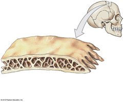
Flat Bones: Thin, parallel surfaces. Provide protection and muscle attachment. Examples: skull roof, sternum, ribs, scapulae.
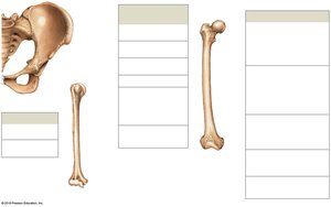
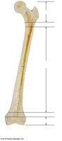
Long Bones: Long and slender, found in limbs, fingers, toes. Example: femur.
Sesamoid Bones: Small, round, flat bones within tendons near joints. Example: patella (kneecap).
Bone Markings (Surface Features)
Bone markings are projections, openings, and depressions that serve as attachment points or passageways for nerves and blood vessels.
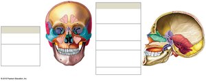
Projections: Sites for muscle, tendon, and ligament attachment; also form joints.
Openings and Depressions: Allow passage of blood vessels and nerves.
Examples of Bone Markings
Process: Projection or bump.
Ramus: Forms an angle with the rest of the bone.
Foramen: Rounded passageway for vessels/nerves.
Fissure: Deep furrow or slit.
Meatus/Canal: Passage or channel.
Trochanter, Crest, Spine, Tubercle, Tuberosity: Various projections for muscle attachment.
Head, Neck, Facet, Condyle, Trochlea: Projections for joint formation.
Sulcus, Fossa: Depressions for vessels or muscle attachment.
Structure of Bones
Long Bones
Diaphysis: Shaft, composed of compact bone, surrounds the medullary cavity.
Epiphysis: Ends of the bone, mostly spongy bone.
Metaphysis: Region where diaphysis and epiphysis meet.
Flat Bones
Consist of spongy bone (diploë) sandwiched between two layers of compact bone.
Bone Tissue
Bone tissue is a dense, supportive connective tissue with specialized cells and a solid matrix.
Matrix: Composed of calcium phosphate (Ca3(PO4)2), calcium hydroxide (Ca(OH)2), forming hydroxyapatite (Ca10(PO4)6(OH)2), and collagen fibers.
Cells: Four types—osteogenic cells, osteoblasts, osteocytes, osteoclasts.
Types of Bone Cells
Osteogenic Cells: Stem cells that produce osteoblasts; found in periosteum and endosteum.
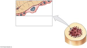
Osteoblasts: Immature cells that synthesize new bone matrix (osteoid).
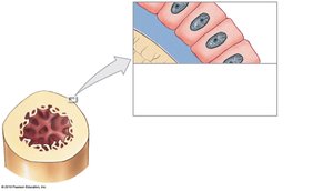
Osteocytes: Mature bone cells maintaining matrix; reside in lacunae, connected by canaliculi.
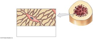
Osteoclasts: Multinucleate cells that resorb bone matrix, important for homeostasis.
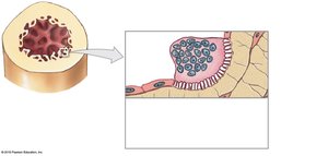
Compact Bone vs. Spongy Bone
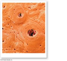
Compact Bone
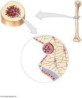
Osteon: Functional unit; concentric lamellae surround a central canal.
Perforating Canals: Carry blood vessels perpendicular to bone surface.
Lamellae: Layers of bone matrix—concentric, interstitial, circumferential.
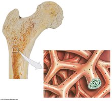
Spongy Bone
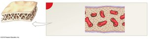
Lacks osteons; matrix forms trabeculae.
Red bone marrow fills spaces, produces blood cells.
Yellow bone marrow stores fat.
Periosteum and Endosteum
Periosteum: Membrane covering bone; outer fibrous and inner cellular layers. Functions in isolation, blood supply, and repair.
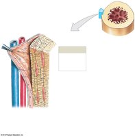
Endosteum: Incomplete cellular layer lining medullary cavity; active in growth and repair.
Bone Formation and Growth
Ossification
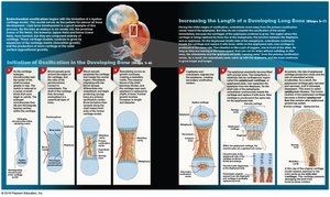
Endochondral Ossification: Most bones form this way; bone replaces hyaline cartilage in seven steps.
Intramembranous Ossification: Occurs in dermis; forms flat bones like mandible and clavicle in five steps.
Bone Growth
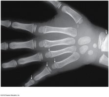
Interstitial Growth: Increases length; involves epiphyseal plate.
Appositional Growth: Increases width; circumferential lamellae added.
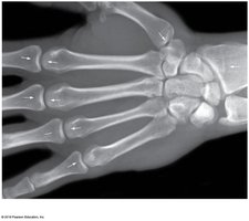
Blood Supply to Bones
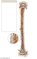
Nutrient Artery and Vein: Enter through nutrient foramina.
Metaphyseal Vessels: Supply epiphyseal cartilages.
Periosteal Vessels: Supply superficial osteons and secondary ossification centers.
Bone Remodeling
Bone remodeling is a continuous process involving osteocytes, osteoblasts, and osteoclasts. It maintains bone integrity by recycling and renewing matrix.
Balanced activity keeps bones healthy.
Imbalance can lead to weakened or strengthened bones.
Exercise, Nutrition, and Hormones
Exercise: Stimulates osteoblasts; bones adapt and become stronger.
Nutrition: Requires calcium, phosphorus, magnesium, fluoride, iron, manganese, vitamins D3, C, A, K, B12.
Hormones: Growth hormone, thyroxine, sex hormones, parathyroid hormone, and calcitonin regulate bone growth and calcium homeostasis.
Calcium Homeostasis
Bones store 99% of the body's calcium. Calcium ions are essential for physiological processes and must be tightly regulated.
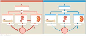
Parathyroid Hormone (PTH): Increases blood calcium by stimulating osteoclasts, increasing intestinal absorption, and reducing kidney excretion.
Calcitonin: Decreases blood calcium by inhibiting osteoclasts, increasing kidney excretion, and reducing intestinal absorption.
Fractures and Repair
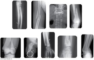
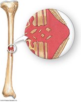
Fractures: Cracks or breaks in bones; classified as open (compound) or closed (simple). Types include transverse, displaced, compression, spiral, epiphyseal, comminuted, greenstick, Colles, and Pott’s fractures.
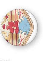
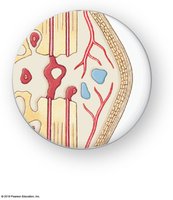
Repair Process: Four steps—fracture hematoma formation, callus formation, spongy bone formation, compact bone formation.
Effects of Aging on the Skeletal System
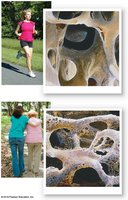
Osteopenia: Reduction in bone mass, begins between ages 30–40; more pronounced in women.
Osteoporosis: Severe bone loss, compromises function; more common in women after menopause.
Cancer: Can cause severe osteoporosis via osteoclast-activating factor.
Summary Table: Bone Cell Types
Cell Type | Function |
|---|---|
Osteogenic cells | Stem cells for osteoblasts; fracture repair |
Osteoblasts | Produce new bone matrix |
Osteocytes | Maintain bone matrix; repair |
Osteoclasts | Resorb bone matrix; release minerals |
Summary Table: Types of Bone Markings
Marking | Purpose |
|---|---|
Process | Projection for muscle attachment |
Foramen | Passage for vessels/nerves |
Fossa | Depression for muscle attachment |
Condyle | Articular surface for joints |
Key Equations
Hydroxyapatite Formation:
Additional info: Academic context was added to clarify bone cell functions, bone markings, and the effects of hormones and nutrition on bone health.
