 Indietro
Bones and Osseous Tissue: Structure, Function, and Development
Guida di studio - Note intelligenti
 Appunti personalizzati basati sui tuoi materiali, ampliati con definizioni chiave, esempi e contesto.
Ch. 6: Bones and Osseous Tissue
Introduction to Bone and Osseous Tissue
Bones are dynamic organs composed of osseous tissue, which provide structural support, protection, and play a central role in mineral homeostasis and blood cell formation. Understanding bone structure and function is essential for comprehending the skeletal system's role in human physiology.
Functions of the Skeleton
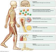
Protection: Bones protect vital organs such as the brain (skull), heart and lungs (rib cage).
Mineral Storage: Bones store essential minerals, primarily calcium and phosphate, which can be released into the bloodstream as needed.
Hematopoiesis: The process of blood cell formation occurs in the red bone marrow.
Movement: Bones act as levers for muscles, facilitating movement.
Support: The skeleton provides the framework that supports the body’s weight.
Fat Storage: Yellow marrow stores triglycerides as an energy reserve.
Hormone Production: Bones produce hormones such as osteocalcin, which influences bone mineralization and metabolism.
Classification of Bones by Shape
Bones are classified based on their shapes, which relate to their functions:
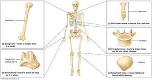
Long Bones: Longer than they are wide (e.g., humerus, femur).
Short Bones: Approximately equal in length and width (e.g., carpals, tarsals).
Flat Bones: Thin, flattened, and often curved (e.g., sternum, ribs, skull bones).
Irregular Bones: Complex shapes that do not fit other categories (e.g., vertebrae).
Sesamoid Bones: Small, round bones embedded within tendons (e.g., patella).
Long Bone Structure
Long bones have a specialized structure that supports their function in movement and weight-bearing:
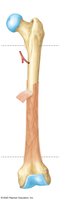
Epiphysis: The expanded ends of the bone, covered with articular (hyaline) cartilage for joint movement.
Diaphysis: The shaft of the bone, composed mainly of compact bone surrounding the medullary cavity.
Periosteum: A dense connective tissue membrane covering the outer surface, containing osteogenic cells for growth and repair.
Endosteum: A thin membrane lining the medullary cavity and trabeculae of spongy bone.
Medullary Cavity: Central cavity containing bone marrow (red or yellow, depending on age).
Epiphyseal Line/Plate: The growth plate, a region of hyaline cartilage where bone growth occurs in children and adolescents.
Compact (Cortical) Bone: Dense outer layer providing strength.
Cancellous (Spongy/Trabecular) Bone: Inner latticework structure containing marrow.
Other Bone Structures
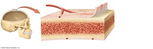
Periosteum: Contains an outer fibrous layer and an inner osteogenic layer; collagen fibers (Sharpey's fibers) anchor it to bone.
Cancellous Bone (Diploë): Found in flat bones, composed of trabeculae and red marrow.
Compact Bone: Forms the hard outer shell of bones.
Bone Marrow
Bone marrow is a soft tissue found within bone cavities and is classified as red or yellow:
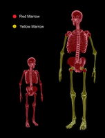
Red Marrow: Site of hematopoiesis (blood cell formation); highly vascularized and found mainly in flat bones and the ends of long bones in adults.
Yellow Marrow: Stores fat; found in the medullary cavity of long bones and increases with age.
Age-Related Changes: Red marrow is gradually replaced by yellow marrow as individuals age.
Bone Composition
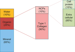
Bones are composed of organic and inorganic components that provide strength and flexibility:
Inorganic Matrix: Mainly hydroxyapatite crystals (calcium phosphate), providing hardness and strength.
Organic Matrix (Osteoid): Primarily type I collagen and non-collagenous proteins (e.g., osteocalcin), providing flexibility and tensile strength.
Glycosaminoglycans (GAGs): Negatively charged sugar chains that contribute to the ground substance.
Bone Cells
Bone tissue contains several specialized cell types, each with distinct functions:
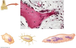
Osteogenic Cells: Stem cells in the periosteum and endosteum that differentiate into osteoblasts; crucial for bone growth and fracture repair.
Osteoblasts: Bone-forming cells that secrete collagen, osteocalcin, and initiate mineralization of the osteoid.
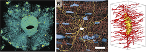
Osteocytes: Mature osteoblasts trapped in lacunae; maintain bone matrix and sense mechanical strain via canaliculi.
Osteoclasts: Large, multinucleated cells derived from macrophages; responsible for bone resorption by releasing acids and enzymes.
Osteocytes and Bone Strain
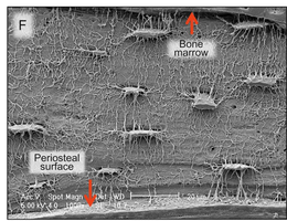
Osteocytes are the most abundant bone cells and play a key role in mechanosensation and bone remodeling:
Reside in lacunae and extend dendritic processes through canaliculi for communication and nutrient exchange.
Sensitive to mechanical strain; signal for bone remodeling when strain is detected.
Structure of Compact and Spongy Bone
Bone tissue is organized into compact and spongy forms, each with unique structural features:
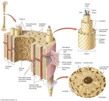
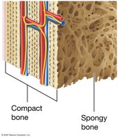
Compact Bone: Composed of osteons (Haversian systems) with concentric lamellae surrounding a central canal containing blood vessels and nerves.
Spongy Bone (Cancellous/Trabecular): Consists of a network of trabeculae; spaces are filled with marrow and lack osteons.
Osteogenesis (Bone Formation)
Bone develops through two primary processes:
Intramembranous Ossification: Occurs within a mesenchymal membrane; forms flat bones like the skull and clavicles. Mesenchymal cells differentiate into osteoblasts, which secrete osteoid and form trabeculae.
Endochondral Ossification: Occurs within a hyaline cartilage scaffold; forms most bones of the body. Chondrocytes create a cartilage model, which is gradually replaced by bone as blood vessels invade and osteoblasts differentiate.
Intramembranous Ossification
Begins in the center of the membrane and moves outward.
Osteoblasts secrete osteoid, forming trabeculae; periosteum develops on the surface.
Endochondral Ossification
Begins with a cartilage model; chondrocytes hypertrophy and die, creating space for bone formation.
Blood vessels invade, bringing osteogenic cells that differentiate into osteoblasts.
Primary ossification center forms in the diaphysis; secondary centers form in the epiphyses.
Growth continues at the epiphyseal plate until adulthood.
Post-Embryonic Bone Growth
Lengthwise Growth (Longitudinal)
Occurs at the epiphyseal plate through endochondral ossification.
Chondrocytes proliferate, hypertrophy, die, and are replaced by bone tissue.
Widthwise Growth (Appositional)
Bone tissue is added to the periosteal surface and removed from the endosteal surface, increasing diameter while maintaining shape.
Bone Remodeling
Bone is continuously remodeled throughout life to repair damage, adapt to mechanical stress (Wolff’s Law), and regulate calcium and phosphate balance:
Osteoclasts resorb bone tissue, while osteoblasts deposit new bone.
Remodeling replaces primary (woven) bone with secondary (lamellar) bone.
Osteocytes detect strain and signal for remodeling.
Calcium Homeostasis
Bone acts as a reservoir for calcium and phosphate, essential for muscle contraction, nerve function, and other physiological processes:
Calcitonin (from the thyroid) stimulates osteoblasts to form bone when blood calcium is high.
Parathyroid hormone (PTH) stimulates osteoclasts to resorb bone when blood calcium is low.
Vitamin D (Calcitriol) increases calcium absorption from the digestive tract and kidneys.
Bone Fractures and Repair
Nondisplaced: Bone ends remain aligned.
Displaced: Bone ends are out of alignment.
Comminuted: Bone is broken into three or more pieces.
Greenstick: Bone is bent and partially broken (common in children).
Fracture repair involves formation of a callus, followed by both endochondral and intramembranous ossification to restore bone integrity.
Summary Table: Bone Cell Types and Functions
Cell Type | Origin | Function |
|---|---|---|
Osteogenic Cell | Mesenchymal stem cell | Differentiate into osteoblasts |
Osteoblast | Osteogenic cell | Form bone matrix |
Osteocyte | Osteoblast | Maintain bone matrix, sense strain |
Osteoclast | Hematopoietic stem cell (macrophage lineage) | Resorb bone |
