Indietro
Cell Cycle, Mitosis, and Protein Synthesis: Study Notes for Anatomy & Physiology
Guida di studio - Note intelligenti
 Appunti personalizzati basati sui tuoi materiali, ampliati con definizioni chiave, esempi e contesto.
Cells: The Living Units
Overview of the Cell Cycle
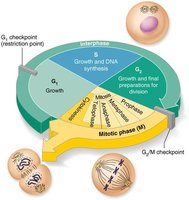
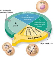
The cell cycle is a series of events that cells go through as they grow and divide. It is essential for growth, tissue repair, and maintenance in multicellular organisms. The cell cycle consists of two major periods: interphase (cell growth and normal function) and cell division (mitotic phase).
Interphase: The cell grows, carries out normal functions, and prepares for division.
Mitotic (M) phase: The cell divides into two daughter cells through mitosis and cytokinesis.
Phases of Interphase
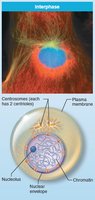
Interphase is the period between cell formation and division, during which the cell performs its normal activities and prepares for division. It is divided into three subphases:
G1 (Gap 1): Vigorous growth and metabolism occur. Cells that permanently stop dividing enter the G0 phase.
S (Synthesis): DNA replication takes place, ensuring each daughter cell receives an identical set of chromosomes.
G2 (Gap 2): Final preparations for cell division are made.
DNA Replication
Before a cell divides, it must replicate its DNA so that each daughter cell receives a complete set of genetic instructions. The process involves:
Unwinding and unzipping of double-stranded DNA helices.
Formation of a replication fork (where strands separate) and a replication bubble (active area of replication).
Each original strand serves as a template for a new complementary strand.
RNA primers initiate DNA synthesis.
Cell Division: Mitosis and Cytokinesis
Overview of Mitosis
Mitosis is the process of nuclear division, ensuring that each daughter cell receives an identical set of chromosomes. It consists of four main stages:
Prophase
Metaphase
Anaphase
Telophase
Cytokinesis is the division of the cytoplasm, resulting in two separate daughter cells.
Stages of Mitosis
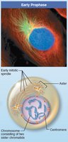
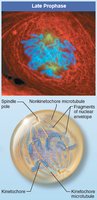
Prophase: Chromatin condenses into visible chromosomes, each consisting of two sister chromatids joined at a centromere. The mitotic spindle forms, and the nuclear envelope breaks down.
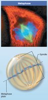
Metaphase: Chromosomes align at the cell's equator (metaphase plate).
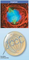
Anaphase: Centromeres split, and sister chromatids (now individual chromosomes) are pulled to opposite poles.
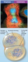
Telophase: Chromosomes uncoil into chromatin, nuclear envelopes reform, nucleoli reappear, and the spindle apparatus disappears.
Cytokinesis: The cytoplasm divides, forming two genetically identical daughter cells.
Control of Cell Division
Cell division is tightly regulated by internal and external signals to ensure proper growth and tissue maintenance.
Go signals: Include critical surface-to-volume ratio and growth factors.
Stop signals: Include contact inhibition (cells stop dividing when they touch each other) and lack of space.
Regulatory proteins: Cyclins and cyclin-dependent kinases (Cdks) regulate progression through the cell cycle.
Checkpoints: Key control points (e.g., G1 checkpoint) ensure the cell is ready for the next phase. If not, the cell may enter G0 (a non-dividing state).
Protein Synthesis
Genetic Code and Genes
DNA serves as the master blueprint for protein synthesis. A gene is a segment of DNA that codes for a specific polypeptide. The genetic code is based on the sequence of nitrogenous bases (adenine, guanine, cytosine, thymine) in DNA.
The code is read in triplets (three bases), each specifying an amino acid.
Genes contain exons (coding regions) and introns (noncoding regions).
The Role of RNA
RNA acts as the intermediary between DNA and protein synthesis. It is synthesized in the nucleus and differs from DNA by having ribose sugar and uracil instead of thymine. There are three main types of RNA:
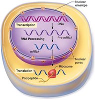
Messenger RNA (mRNA): Carries the genetic code from DNA to ribosomes (transcription).
Ribosomal RNA (rRNA): Structural component of ribosomes, facilitates translation.
Transfer RNA (tRNA): Brings amino acids to the ribosome, matching codons with anticodons (translation).
Steps of Protein Synthesis
Transcription: DNA information is copied into mRNA in the nucleus.
Translation: mRNA is decoded at the ribosome to assemble a polypeptide chain.
Cellular Maintenance and Death
Autophagy
Autophagy is the process by which cells remove damaged organelles and cytoplasmic debris. Autophagosomes engulf the material, which is then degraded by lysosomes.
Ubiquitin-Proteasome Pathway
Proteins that are damaged or no longer needed are tagged with ubiquitin and degraded by proteasomes, recycling amino acids and ubiquitin for reuse.
Apoptosis
Apoptosis is programmed cell death, a controlled process that eliminates damaged, infected, or unnecessary cells. It involves activation of caspases, degradation of DNA and cytoskeleton, and phagocytosis of cell remnants by macrophages.
Cell Division, Aging, and Disease
Cell Division in Growth and Repair
Cell division is essential for growth, replacing short-lived cells, and repairing tissues. Hyperplasia is increased cell production, while atrophy is a decrease in cell size or number due to reduced stimulation or use.
Cell Aging
Several theories explain cell aging:
Wear and tear theory: Accumulated damage from chemicals and free radicals.
Mitochondrial theory: Free radicals impair mitochondrial function.
Immune system theory: Autoimmune responses and declining immunity.
Genetic theory: Programmed limits on cell division, involving telomeres (protective DNA sequences at chromosome ends) and telomerase (an enzyme that extends telomeres, active in germ cells and cancer cells).
Clinical Example: Progeria
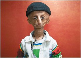
Progeria is a rare genetic disorder that mimics accelerated aging. It is caused by a defective protein in the nuclear lamina, leading to an unstable nucleus. Symptoms include slow growth, thinning hair, brittle bones, arthritis, and severe cardiovascular disease. Life expectancy is typically around 20 years. Treatments that stimulate autophagy may help clear defective proteins.