Indietro
Cell Structures and Intercellular Junctions: Study Notes for Anatomy & Physiology
Guida di studio - Note intelligenti
 Appunti personalizzati basati sui tuoi materiali, ampliati con definizioni chiave, esempi e contesto.
4.3 Eukaryotic Cells
Introduction to Eukaryotic Cells
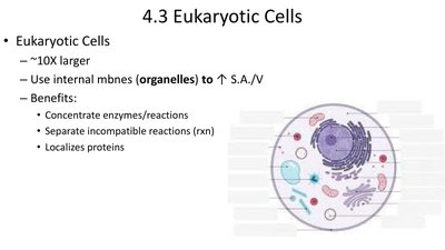
Eukaryotic cells are complex cells characterized by the presence of membrane-bound organelles. They are typically about ten times larger than prokaryotic cells and use internal membranes (organelles) to increase surface area-to-volume ratio, which enhances cellular efficiency.
Benefits of organelles: Concentrate enzymes and reactions, separate incompatible reactions, and localize proteins for specific functions.
Eukaryotic Cell Structures and Functions
Nucleus
The nucleus is the control center of the cell, containing genetic material and coordinating activities such as growth, metabolism, and protein synthesis.
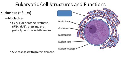
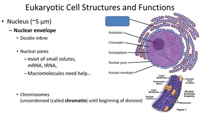
Nucleolus: Site for ribosome synthesis, including rRNA, tRNA, proteins, and partially constructed ribosomes. The size of the nucleolus changes with protein demand.
Nuclear envelope: Double membrane structure with nuclear pores that regulate the movement of small solutes, mRNA, and tRNA. Macromolecules require assistance to pass through.
Chromosomes: Uncondensed chromosomes are called chromatin until cell division begins.
Ribosomes
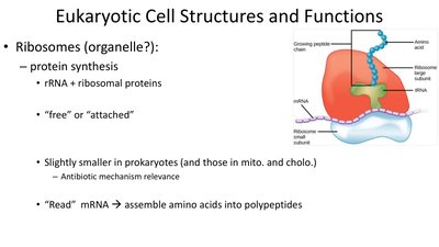
Ribosomes are molecular machines responsible for protein synthesis. They are composed of rRNA and ribosomal proteins and can be found free in the cytoplasm or attached to the endoplasmic reticulum.
Function: Read mRNA and assemble amino acids into polypeptides.
Prokaryotic vs. Eukaryotic Ribosomes: Ribosomes are slightly smaller in prokaryotes and in mitochondria/chloroplasts, which is relevant for antibiotic mechanisms.
Eukaryotic Plasma Membrane
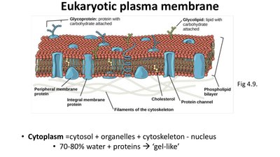
The plasma membrane is a selectively permeable barrier composed of a phospholipid bilayer with embedded proteins, glycoproteins, and glycolipids. It separates the cell from its environment and regulates the movement of substances in and out.
Cytoplasm: Includes cytosol, organelles, cytoskeleton, and excludes the nucleus. It is 70-80% water with proteins, giving it a gel-like consistency.
4.4 Endomembrane System (ES)
Overview of the Endomembrane System
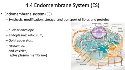
The endomembrane system is a network of membranes within eukaryotic cells that synthesizes, modifies, stores, and transports lipids and proteins. It includes the nuclear envelope, endoplasmic reticulum, Golgi apparatus, lysosomes, and vesicles.
Endoplasmic Reticulum (ER)
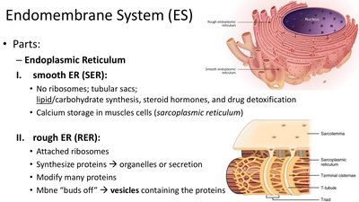
Smooth ER (SER): Lacks ribosomes; involved in lipid and carbohydrate synthesis, steroid hormone production, and detoxification. In muscle cells, it stores calcium (sarcoplasmic reticulum).
Rough ER (RER): Studded with ribosomes; synthesizes proteins for organelles or secretion, modifies proteins, and forms vesicles containing proteins.
Golgi Apparatus
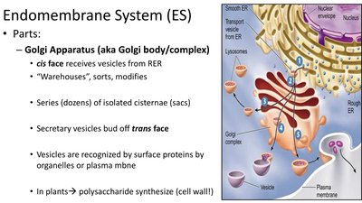
The Golgi apparatus (Golgi body/complex) is a series of flattened sacs that function as the cell's "warehouse," sorting, modifying, and packaging proteins and lipids for delivery. The cis face receives vesicles from the RER, and the trans face releases secretory vesicles.
In plants, the Golgi synthesizes polysaccharides for the cell wall.
Lysosomes
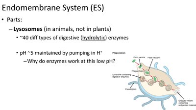
Lysosomes are membrane-bound organelles in animal cells containing about 40 types of digestive (hydrolytic) enzymes. They maintain an acidic pH (~5) by pumping in H+ ions, which is optimal for enzyme activity.
Function: Breakdown of macromolecules, cellular debris, and foreign substances.
Vacuoles
Vacuoles are large, membrane-bound sacs found in plants, fungi, and some protists. Their contents and functions vary:
Food vacuole: Stores nutrients.
Contractile vacuole: Expels excess water.
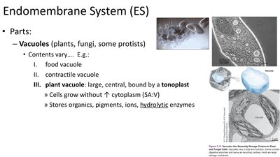
Plant vacuole: Large, central, and bound by a tonoplast; stores organics, pigments, ions, and hydrolytic enzymes. It helps cells grow without increasing cytoplasm volume and maintains cell rigidity.
Peroxisomes
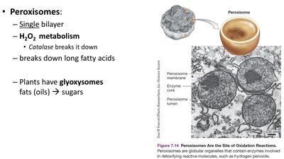
Peroxisomes are single-membrane organelles involved in the metabolism of hydrogen peroxide (H2O2). The enzyme catalase breaks down H2O2 and peroxisomes also break down long fatty acids. In plants, glyoxysomes convert fats (oils) to sugars.
Energy Generating Organelles
Chloroplasts
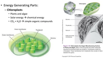
Chloroplasts are found in plants and algae and are responsible for photosynthesis, converting solar energy into chemical energy. They use CO2 and H2O to produce simple organic compounds.
Mitochondria
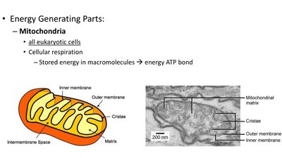
Mitochondria are present in all eukaryotic cells and are the site of cellular respiration. They convert stored energy in macromolecules into ATP, the cell's energy currency.
4.5 Dynamic Cytoskeleton
Structure and Function of the Cytoskeleton
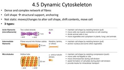
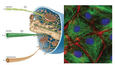
The cytoskeleton is a dense, complex network of fibers that provides structural support, anchors organelles, and enables cell movement and shape changes. It is dynamic, constantly reorganizing to meet cellular needs.
Actin filaments (microfilaments): Maintain cell shape by resisting tension (pull), involved in cell movement and division, and move organelles and cytoplasm.
Intermediate filaments: Provide structural stability, anchor organelles, and resist tension. Examples include keratins and lamins.
Microtubules: Hollow tubes that maintain cell shape by resisting compression (push), move chromosomes during cell division, and provide tracks for intracellular transport.
Intercellular Junctions
Gap Junctions
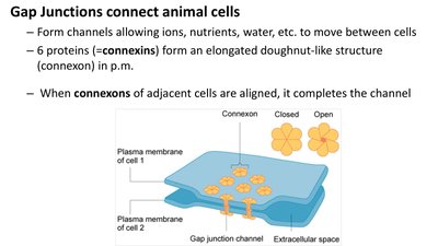
Gap junctions connect animal cells by forming channels that allow ions, nutrients, water, and other molecules to move between cells. Six proteins called connexins form a doughnut-like structure (connexon) in the plasma membrane. When connexons of adjacent cells align, they complete the channel.
Desmosomes
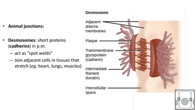
Desmosomes are short proteins (cadherins) in the plasma membrane that act as "spot welds" to join adjacent cells in tissues that stretch, such as the heart, lungs, and muscles. They provide mechanical stability by anchoring intermediate filaments.
Tight Junctions
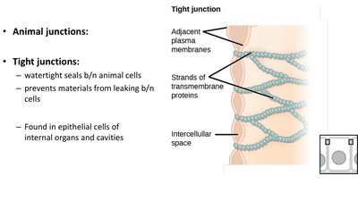
Tight junctions create watertight seals between animal cells, preventing materials from leaking between cells. They are commonly found in epithelial cells of internal organs and cavities.
Intercellular Junctions in Plants
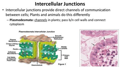
Intercellular junctions provide direct channels of communication between cells. In plants, plasmodesmata are channels that pass through cell walls and connect the cytoplasm of adjacent cells, allowing the exchange of molecules and ions.
Extracellular Structures
Plant Cell Wall
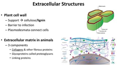
The plant cell wall provides structural support, primarily through cellulose and lignin, and acts as a barrier to infection. Plasmodesmata connect plant cells through the cell wall.
Extracellular Matrix in Animals
The extracellular matrix (ECM) in animals is composed of three main components: collagens and other fibrous proteins, glycoproteins called proteoglycans, and linking proteins. The ECM provides structural support, mediates cell signaling, and anchors cells within tissues.
Prokaryotic Surface Structures
Plasma Membrane and Cell Wall
Prokaryotic cells have a plasma membrane composed of a lipid bilayer, with differences in lipid and protein composition compared to eukaryotes. Most prokaryotes have a cell capsule or wall for shape, protection, and attachment.
Fibrous composite: Cross-linked network of long filaments; some have a glycocalyx (capsule or slime layer).
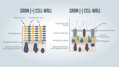
Gram-Positive vs. Gram-Negative Cell Walls
Prokaryotic cell walls can be classified as Gram-positive or Gram-negative based on their structure and staining properties.
Feature | Gram-Positive | Gram-Negative |
|---|---|---|
Peptidoglycan Layer | Thick | Thin |
Teichoic Acids | Present | Absent |
Outer Membrane | Absent | Present |
Lipopolysaccharide | Absent | Present |
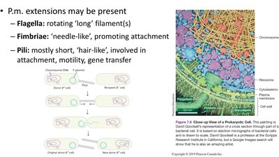
Plasma Membrane Extensions
Some prokaryotes have plasma membrane extensions such as flagella (for motility), fimbriae (for attachment), and pili (for attachment, motility, and gene transfer).
Putting the Parts into a Whole
Cellular Structures and Function
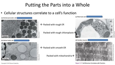
The structure of cellular components correlates with their function. For example, animal pancreatic cells are packed with rough ER for protein synthesis, plant leaf cells with chloroplasts for photosynthesis, animal testis cells with smooth ER for lipid synthesis, and cardiac muscle cells with mitochondria for energy production.