 Indietro
Cells and Tissues: Structure and Function in Anatomy & Physiology
Guida di studio - Note intelligenti
 Appunti personalizzati basati sui tuoi materiali, ampliati con definizioni chiave, esempi e contesto.
Cells and Tissues
Introduction to Cells
Cells are the fundamental structural and functional units of all living organisms. The human body contains between 50 to 100 trillion cells, each varying in shape, size, organelle content, and function. The design of a cell is closely related to its specific function.
Key Point 1: Cells are diverse in structure and function, reflecting their specialized roles in the body.
Key Point 2: The basic parts of a cell include the nucleus, plasma membrane, and cytoplasm.
Example: Nerve cells are long to transmit signals, while red blood cells are biconcave for gas exchange.
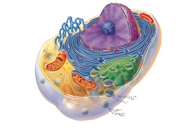
Anatomy of a Generalized Cell
Basic Cell Components
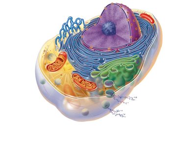
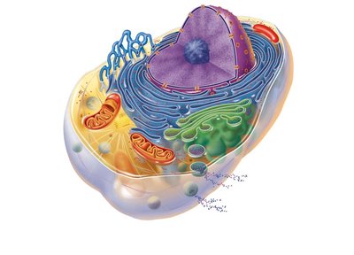
A generalized cell model includes many known cell structures, though no single cell contains all possible organelles. The three main parts are:
Nucleus: Contains DNA and controls cell activities.
Plasma Membrane: Defines the cell boundary and regulates entry/exit of substances.
Cytoplasm: Gel-like fluid between nucleus and membrane, containing organelles and site of most cellular activities.
The Nucleus
Structure and Function
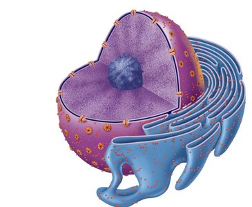
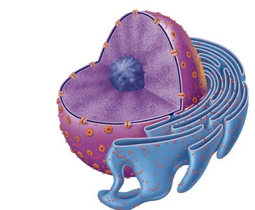
The nucleus is the control center of the cell, housing genetic material (DNA) necessary for protein synthesis and cell reproduction. It consists of three regions:
Nuclear Envelope: Double membrane enclosing the nucleus, with nuclear pores for material exchange.
Nucleolus: Site of ribosome assembly; ribosomes exit through nuclear pores to participate in protein synthesis.
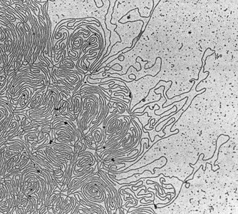
Chromatin: Loosely coiled DNA and protein, present when the cell is not dividing; condenses into chromosomes during cell division.
Plasma Membrane
Structure and Function
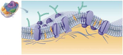
The plasma membrane acts as a barrier separating cellular contents from the external environment. It is selectively permeable, regulating what enters and leaves the cell, and is involved in signal transduction and cell adhesion.
Phospholipid Bilayer: Composed of hydrophilic heads facing outward and hydrophobic tails inward, forming a double layer.
Cholesterol: Stabilizes the membrane and reduces permeability to water-soluble substances.
Proteins: Provide functionality such as receptors, channels, transporters, enzymes, and cell recognition.
Sugars: Glycolipids and glycoproteins form the glycocalyx, aiding in cell recognition and adhesion.
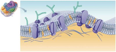
Cell part | Structure | Functions |
|---|---|---|
Plasma Membrane | Double layer of lipids (phospholipids, cholesterol, proteins, sugars) | Acts as cell barrier, regulates transport, maintains internal conditions, cell recognition |
Cytoplasm
Components and Functions
The cytoplasm is the site of most cellular activities and contains three major elements:
Cytosol: Fluid suspending other elements.
Organelles: Specialized compartments performing specific functions.
Inclusions: Stored nutrients or cell products.
Cytoplasmic Organelles
Mitochondria
Mitochondria are double-membrane organelles with inner folds (cristae) containing enzymes for ATP production via cellular respiration. The matrix is the internal fluid.
Function: Major site of ATP production in presence of O2.
Ribosomes
Ribosomes are dense granules composed of ribosomal RNA and protein, serving as the site of protein synthesis. They are found attached to rough ER or free in the cytoplasm.
Endoplasmic Reticulum (ER)
Rough ER: Studded with ribosomes; synthesizes and transports proteins.
Smooth ER: Lacks ribosomes; synthesizes lipids and detoxifies substances.
Protein Synthesis and Transport
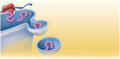
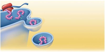
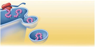
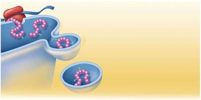
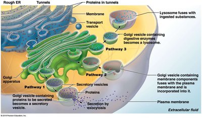
Proteins synthesized on ribosomes attached to rough ER are transported via vesicles to the Golgi apparatus for further processing.
Golgi Apparatus
The Golgi apparatus modifies and packages proteins received from the rough ER, producing secretory vesicles, in-house proteins/lipids, and lysosomes.
Lysosomes and Peroxisomes
Lysosomes: Membrane-bound sacs containing digestive enzymes; digest damaged organelles and foreign particles.
Peroxisomes: Contain oxidase enzymes; detoxify harmful substances and convert free radicals to water.
Cytoskeleton
Structure and Function
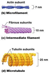
The cytoskeleton is a network of protein structures providing internal support and shape to the cell. It consists of three types:
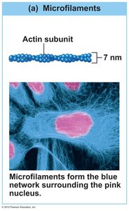
Microfilaments: Composed of actin and myosin; involved in cell shape changes and motility.
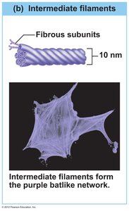
Intermediate Filaments: Provide structural support and help form desmosomes.
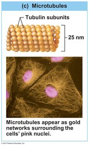
Microtubules: Composed of tubulin; maintain cell shape, distribute organelles, and form mitotic spindle.
Type | Subunit | Diameter | Function |
|---|---|---|---|
Microfilament | Actin | 7 nm | Cell shape, movement |
Intermediate filament | Fibrous protein | 10 nm | Structural support |
Microtubule | Tubulin | 25 nm | Shape, organelle movement, mitosis |
Centrioles
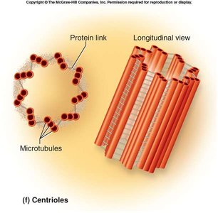
Centrioles are rod-shaped structures made of microtubules, located near the nucleus. They direct the formation of the mitotic spindle during cell division.
Cell Extensions
Types and Functions
Some cells possess surface extensions:
Cilia: Move materials across the cell surface.
Flagella: Propel sperm cells.
Microvilli: Increase surface area for absorption.
Cell Physiology
Cell Functions
Cells perform essential functions such as metabolism, digestion, waste disposal, reproduction, growth, movement, and response to stimuli.
Membrane Transport
Types of Transport
The cell membrane is selectively permeable, allowing certain substances to enter or leave the cell. Transport mechanisms are classified as passive or active:
Passive Transport: Does not require ATP; includes diffusion, facilitated diffusion, osmosis, and filtration.
Active Transport: Requires ATP; includes solute pumping, endocytosis, and exocytosis.
Diffusion
Diffusion is the movement of molecules from high to low concentration, down a concentration gradient, until equilibrium is reached. Simple diffusion occurs for small, lipid-soluble molecules.
Osmosis: Diffusion of water across a selectively permeable membrane.
Facilitated Diffusion: Large or lipid-insoluble substances use carrier or channel proteins.
Filtration: Water and small solutes are forced through a membrane by hydrostatic pressure.
Active Transport
Active transport moves substances against their concentration gradient using carrier proteins and ATP. The sodium-potassium pump is a classic example.
Equation: (energy release for transport)
Vesicular Transport
Exocytosis: Vesicles fuse with the plasma membrane to expel contents.
Endocytosis: Plasma membrane engulfs material, forming a vesicle inside the cell (includes pinocytosis and phagocytosis).
Cell Division
Mitosis and Meiosis
Cell division is essential for growth, repair, and reproduction. Mitosis produces two genetically identical daughter cells, while meiosis produces four gametes with half the chromosome number.
DNA Replication: DNA is duplicated before cell division, ensuring each daughter cell receives a complete set.
Equation: (base pairing during replication)
Body Tissues
Types of Tissues
Tissues are groups of cells similar in structure and function. The four main types are:
Epithelial Tissue: Covers surfaces, lines cavities, forms glands.
Connective Tissue: Supports, binds, and protects body structures.
Muscle Tissue: Contracts to produce movement.
Nervous Tissue: Transmits electrical impulses.
Epithelial Tissue
Special Characteristics: Tightly packed, little extracellular material, apical surface, basement membrane, avascular, regenerates easily.
Classification: By cell arrangement (simple, stratified, pseudostratified) and cell shape (squamous, cuboidal, columnar).
Connective Tissue
Extracellular Matrix: Composed of fibers (collagen, elastic, reticular) and ground substance.
Types: Bone, cartilage, dense regular, areolar, adipose, blood.
Muscle Tissue
Skeletal Muscle: Long, cylindrical, striated, multinucleated, voluntary.
Cardiac Muscle: Striated, short, branched, intercalated discs, involuntary.
Smooth Muscle: Spindle-shaped, no striations, involuntary.
Nervous Tissue
Neurons: Transmit electrical impulses.
Neuroglia: Support and protect neurons.
Additional info: Academic context and explanations have been expanded for clarity and completeness. Only images directly relevant to the adjacent content have been included.
