 Indietro
Central Nervous System: The Brain and Spinal Cord
Guida di studio - Note intelligenti
 Appunti personalizzati basati sui tuoi materiali, ampliati con definizioni chiave, esempi e contesto.
Central Nervous System Overview
The central nervous system (CNS) consists of the brain and spinal cord, serving as the main control center for processing and transmitting information throughout the body. This section explores the structure, protection, and function of the spinal cord and brain, as well as their associated disorders.
Spinal Cord
Functional Anatomy of the Spinal Cord
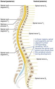
The spinal cord is a cylindrical structure enclosed within the vertebral column. It begins at the foramen magnum of the skull and typically ends at the level of the L1 or L2 vertebra. The spinal cord provides two-way communication between the brain and the body and serves as a major reflex center, where reflexes are initiated and completed.
Conduction: Bundles of axons conduct sensory information up to the brain and motor signals down to muscles and glands.
Reflex Center: Many reflexes are processed directly in the spinal cord without brain involvement.
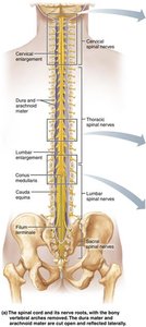
Gross Anatomy and Protection
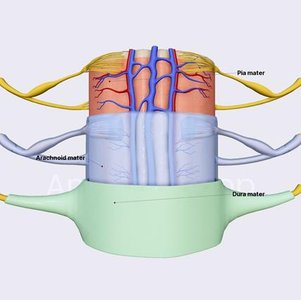
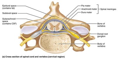
The spinal cord is protected by three main structures: bone (vertebrae), meninges, and cerebrospinal fluid (CSF). The meninges are three connective tissue membranes that surround the CNS:
Dura mater: Outermost, tough membrane against bone.
Arachnoid mater: Middle, web-like membrane.
Pia mater: Innermost, delicate membrane adhering to the spinal cord.
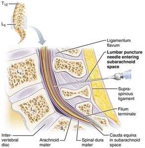
The subarachnoid space, filled with CSF, lies between the arachnoid and pia mater and is the site for lumbar puncture procedures.
Spinal Cord Structure and Features
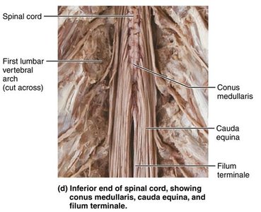
Conus medullaris: The cone-shaped terminal end of the spinal cord.
Filum terminale: A fibrous extension of the pia mater anchoring the spinal cord to the coccyx.
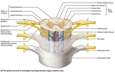
Denticulate ligaments: Extensions of pia mater that secure the cord to the dura mater.
Cervical and lumbar enlargements: Regions where nerves serving the limbs arise.
Cauda equina: A bundle of spinal nerve roots extending below the conus medullaris.
Spinal Nerves and Segmentation
Spinal nerves are part of the peripheral nervous system (PNS) and attach to the spinal cord by 31 paired roots. Each segment of the spinal cord is named for the spinal nerves that arise from it. The lower spinal nerves form the cauda equina.
Cross-Sectional Anatomy
In cross-section, the spinal cord displays a central core of gray matter (containing neuron cell bodies and dendrites) surrounded by white matter (myelinated axons). The central canal runs the length of the cord and is filled with CSF.
Spinal Cord Trauma and Disorders
Types of Functional Loss
Paresthesia: Loss of sensory function due to damage to dorsal roots or sensory tracts.
Paralysis: Loss of motor function due to damage to ventral roots or ventral horn cells.
Flaccid paralysis: Severe damage prevents impulses from reaching muscles; muscles atrophy.
Spastic paralysis: Damage to upper motor neurons; muscles are stimulated by reflex activity but lack voluntary control.
Transection (severing) of the spinal cord results in total loss of motor and sensory function below the injury. The level of injury determines the extent of paralysis:
Paraplegia: Transection between T1 and L1 (loss of lower limb function).
Quadriplegia: Transection in the cervical region (loss of all limb function).
Spinal shock: Temporary loss of function below the lesion.
Amyotrophic Lateral Sclerosis (ALS)
ALS, or Lou Gehrig’s disease, involves the destruction of ventral horn motor neurons and fibers of the pyramidal tract. Symptoms include loss of the ability to speak, swallow, and breathe, with death typically occurring within five years. Causes include genetic mutations and environmental factors affecting RNA processing.
Brain: Structure and Function
Meninges of the Brain
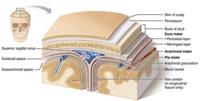
The brain is protected by the same three meninges as the spinal cord. The dura mater forms a fibrous sac and dural sinuses, which collect venous blood. The arachnoid mater contains large blood vessels in the subarachnoid space, and the pia mater is the thinnest, innermost layer.
Cerebral Hemispheres
The cerebral hemispheres form the superior part of the brain and account for 83% of its mass. The surface is marked by gyri (ridges), sulci (shallow grooves), and fissures (deep grooves). Each hemisphere is divided into five lobes: frontal, parietal, temporal, occipital, and insula.
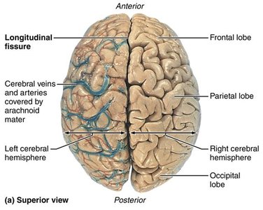
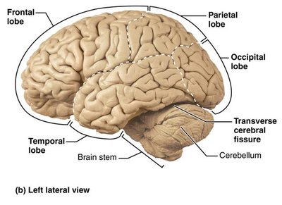
Regions of the Cerebral Hemispheres
Cerebral cortex: The outer layer of gray matter, site of conscious mind functions such as awareness, sensory perception, voluntary movement, communication, memory, and understanding.
Internal white matter: Myelinated axons connecting different brain regions.
Basal nuclei: Deep clusters of gray matter involved in movement regulation and cognition.
Cerebral Cortex: Functional Areas
Motor areas: Control voluntary movement (located in the frontal lobe).
Sensory areas: Provide conscious awareness of sensation (located in parietal, insular, temporal, and occipital lobes).
Association areas: Integrate diverse information, store memories, and link sensations to emotions and actions.
Each hemisphere controls the contralateral side of the body, and some functions are lateralized to one hemisphere. Conscious behavior involves the entire cortex.
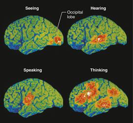
White Matter and Basal Nuclei
White matter: Composed of tracts of myelinated axons, including the corpus callosum, which connects the two hemispheres.
Basal nuclei (ganglia): Influence muscle movements, regulate movement intensity, and filter out inappropriate responses.
Divisions of the Brain
Cerebral hemispheres
Diencephalon: Includes the thalamus, hypothalamus, and pineal gland.
Brain stem: Composed of the midbrain, pons, and medulla oblongata.
Cerebellum: Coordinates movement and balance.
Diencephalon
Thalamus: Relay station for sensory and motor signals to the cortex; sorts, edits, and relays information.
Hypothalamus: Regulates homeostasis, autonomic functions, emotions, body temperature, hunger, thirst, sleep-wake cycles, and endocrine system.
Pineal gland: Secretes melatonin, regulating sleep cycles.
Brain Stem
Midbrain: Contains nuclei for motor control, pain filtering, and visual/auditory reflexes. Degeneration leads to Parkinson's disease.
Pons: Relays signals between the cerebrum and cerebellum; origin of cranial nerves V, VI, and VII.
Medulla oblongata: Autonomic reflex center controlling cardiovascular, respiratory, and other vital functions.
Cerebellum
The cerebellum processes input from the cortex, brain stem, and sensory receptors to coordinate precise movements and maintain balance. It is highly active during tasks requiring timing and prediction.
Functional Brain Systems
Limbic system: Involved in emotion, memory, and motivation; interacts with the hypothalamus and prefrontal cortex.
Reticular formation: Maintains consciousness and alertness; filters sensory input and regulates visceral functions.
Cerebrospinal Fluid (CSF) and Ventricles
CSF: Produced by ependymal cells lining the ventricles; provides buoyancy, chemical stability, and waste removal for the brain.
Ventricles: Fluid-filled chambers within the brain, continuous with the central canal of the spinal cord and filled with CSF.
Blood Supply and Blood Brain Barrier
The blood brain barrier maintains a stable environment for the brain by regulating the movement of substances from the blood into neural tissue. It is formed by tight junctions between endothelial cells, supported by astrocytes. Lipid-soluble substances pass freely, while others require specific transport mechanisms. The barrier is absent in some brain regions, such as the hypothalamus.
Summary Table: Spinal Cord and Brain Protection
Structure | Function | Location |
|---|---|---|
Dura mater | Outermost, tough protection | Against bone |
Arachnoid mater | Middle, web-like, contains CSF | Between dura and pia mater |
Pia mater | Innermost, delicate, adheres to CNS | Directly on brain/spinal cord |
CSF | Cushions, nourishes, removes waste | Subarachnoid space, ventricles |
Additional info: The notes above expand on the original content by providing definitions, examples, and additional academic context for clarity and completeness.
