 Indietro
Chapter 21: The Immune System – Innate and Adaptive Body Defenses
Guida di studio - Note intelligenti
 Appunti personalizzati basati sui tuoi materiali, ampliati con definizioni chiave, esempi e contesto.
The Immune System: Overview
Introduction to Immune System
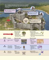
The immune system is a functional system that provides resistance to disease-causing microorganisms such as bacteria, fungi, and viruses. It is not an anatomical organ system but consists of a variety of molecules and immune cells, especially lymphocytes, that reside in lymphoid tissues and circulate in body fluids. The immune system operates through three lines of defense, analogous to a medieval castle's protective layers.
First Line of Defense: Surface barriers (skin and mucosae)
Second Line of Defense: Innate internal defenses (inflammation, antimicrobial proteins, phagocytes)
Third Line of Defense: Adaptive (specific) defense system (lymphocytes and antibodies)
Innate Defenses
Surface Barriers: First Line of Defense
Surface barriers include the skin and mucous membranes, along with their secretions. These barriers are present at birth and are highly effective at preventing the entry of most microbes.
Keratinized Epidermis: Keratin is resistant to weak acids, bases, bacterial enzymes, and toxins.
Mucosae: Provide mechanical barriers within the body, lining tracts.
Protective Chemicals:
Acid Mantle: Acidity of skin, vaginal, and stomach secretions inhibits bacterial growth.
Enzymes: Lysozyme in saliva, mucus, and lacrimal fluid kills microbes; stomach enzymes digest pathogens.
Mucin: Sticky mucus traps microbes in digestive and respiratory tracts.
Defensins: Antimicrobial peptides secreted in response to barrier breach.
Other Chemicals: Lipids in sebum and dermcidin in sweat are toxic to bacteria.
Respiratory Tract Modifications: Mucus-coated hairs in nose trap particles; cilia sweep mucus toward mouth.
Innate Internal Defenses: Second Line of Defense
If surface barriers are breached, innate internal defenses are activated. These include nonspecific cellular and chemical means to protect the body, such as phagocytes, natural killer cells, antimicrobial proteins, inflammation, and fever.
Pattern Recognition Receptors: Identify pathogens by recognizing specific-shaped molecules not found on human cells.
Toll-like Receptors (TLRs): Central role in triggering immune responses; humans have 11 TLRs, each recognizing a particular class of microbe.
Phagocytes
Phagocytes are white blood cells that ingest and digest foreign invaders and cellular debris. The main types are neutrophils and macrophages.
Neutrophils: Most abundant; phagocytize infectious material.
Macrophages: Most voracious; can be free (wandering) or fixed (resident in organs).
Phagocytosis Process
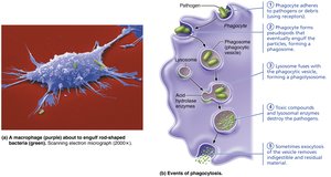
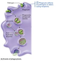
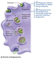
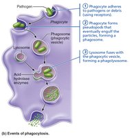
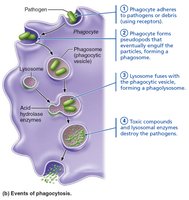
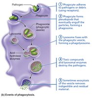
Phagocyte receptors bind to particle, which is pulled inside and enclosed in a phagosome.
Phagosome fuses with lysosome to form a phagolysosome.
Lysosomal enzymes digest contents; some pathogens resist digestion.
Helper T cells can stimulate macrophages to produce a respiratory burst, releasing free radicals and oxidizing chemicals to kill pathogens.
Defensins from neutrophils pierce pathogen membranes.
Natural Killer (NK) Cells
NK cells are large granular lymphocytes that police blood and lymph, targeting cells with abnormal surface proteins (e.g., lack of MHC). They induce apoptosis in cancer and virus-infected cells and secrete chemicals to enhance inflammation.
Inflammation: Tissue Response to Injury
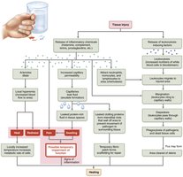
Inflammation is a nonspecific response to tissue injury, preventing spread of pathogens, disposing of debris, alerting the adaptive immune system, and setting the stage for repair. The four cardinal signs are redness, heat, swelling, and pain.
Inflammatory Chemical Release: Histamine, kinins, prostaglandins, cytokines, and complement proteins.
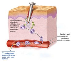
Vasodilation and Increased Permeability: Local hyperemia and exudate formation.
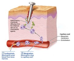
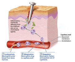
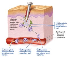
Phagocyte Mobilization: Steps include leukocytosis, margination, diapedesis, and chemotaxis.
Antimicrobial Proteins
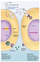
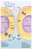
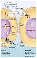
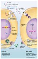
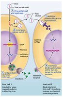
Antimicrobial proteins enhance innate defenses by attacking microbes directly or hindering their reproduction. The most important are interferons and complement proteins.
Interferons: Immune-modulating proteins that interfere with viral replication and activate NK cells and macrophages.
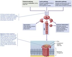
Complement System: At least 20 plasma proteins that amplify inflammation, promote phagocytosis, and cause cell lysis.
Adaptive Defenses
Adaptive Immune System: Third Line of Defense
The adaptive immune system is a specific defensive system that targets and eliminates almost any pathogen. It consists of two overlapping arms:
Humoral Immunity: Antibody-mediated, targets extracellular pathogens.
Cellular Immunity: Cell-mediated, targets infected, cancerous, or foreign cells.
Antigens
Antigens are substances that trigger the body's adaptive defenses. They are usually large, complex molecules that are foreign to the body.
Complete Antigens: Have immunogenicity and reactivity.
Haptens: Incomplete antigens that become immunogenic when attached to a protein carrier.
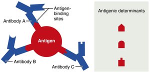
Antigenic Determinants: Parts of antigen that antibodies or lymphocyte receptors bind to.
Self-Antigens: MHC Proteins
Self-antigens are proteins on body cell surfaces that are not antigenic to self but are to others. MHC proteins display peptides for T lymphocytes to recognize.
B and T Lymphocytes and Antigen-Presenting Cells
Three crucial cell types are involved in adaptive immunity:
B Lymphocytes: Provide humoral immunity.
T Lymphocytes: Provide cellular immunity.
Antigen-Presenting Cells (APCs): Present antigens to T cells.
Lymphocyte Development, Maturation, and Activation
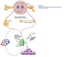
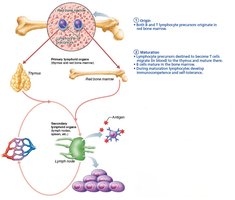
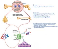
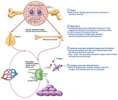
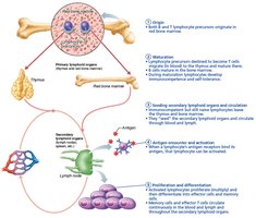
Origin: Hematopoietic stem cells in red bone marrow.
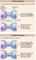
Maturation: Occurs in bone marrow (B cells) or thymus (T cells); involves immunocompetence and self-tolerance.
Seeding Secondary Organs: Naive cells colonize lymph nodes, spleen, etc.
Antigen Encounter and Activation: Clonal selection and activation.
Proliferation and Differentiation: Formation of effector and memory cells.
Humoral Immunity
Activation and Differentiation of B Cells
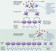
Naive B cells are activated by antigen binding, followed by endocytosis and proliferation into plasma cells (antibody-secreting) and memory cells.
Immunological Memory
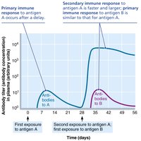
Primary Response: Lag period of 3–6 days; plasma antibody levels peak in 10 days.
Secondary Response: Faster, stronger, and longer-lasting due to memory cells.
Active and Passive Humoral Immunity
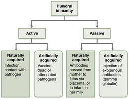
Active Immunity: B cells produce antibodies; can be naturally (infection) or artificially (vaccination) acquired.
Passive Immunity: Antibodies are introduced; no memory cells formed; protection is temporary.
Antibodies (Immunoglobulins)
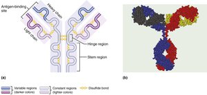
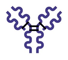
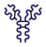
Antibodies are proteins secreted by plasma cells, grouped into five classes: IgG, IgA, IgM, IgE, and IgD. They bind specifically to antigens and tag them for destruction.
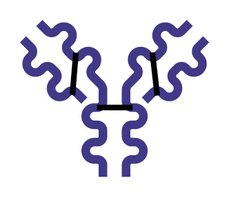
Structure: Four polypeptide chains (two heavy, two light) with variable and constant regions.
Effector Functions: Activate complement, circulate in blood, cross placenta, found in secretions.
Antibody Classes
Class | Structure | Main Function |
|---|---|---|
IgG | Monomer | Main antibody in secondary response; crosses placenta |
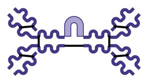
IgA | Dimer/Monomer | Found in secretions; prevents pathogen entry |
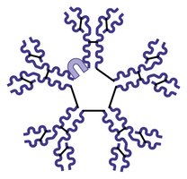
IgM | Pentamer | First antibody released; potent agglutinating agent |
IgE | Monomer | Triggers histamine release; involved in allergies |
IgD | Monomer | B cell surface receptor |
Antibody Functions
Neutralization: Block binding sites on pathogens/toxins.
Agglutination: Clumping of antigens.
Precipitation: Formation of insoluble complexes.
Complement Activation: Leads to cell lysis.
Cellular Immunity
T Lymphocytes: Directing Adaptive Immunity
T cells provoke a cellular immune response when presented with antigens. CD4 cells become helper or regulatory T cells; CD8 cells become cytotoxic T cells.
MHC Proteins and Antigen Presentation
T cells respond only to processed fragments of antigens displayed by MHC proteins. MHC restriction ensures CD4 and CD8 cells bind antigens only on specific MHC classes.
MHC Class | Cell Type | Antigen Source | Role |
|---|---|---|---|
I | All body cells except RBCs | Endogenous | Activates CD8 cells |
II | APCs (dendritic, macrophages, B cells) | Exogenous | Activates CD4 cells |
Activation and Differentiation of T Cells
T cell activation requires antigen binding and co-stimulation. Activated T cells proliferate and differentiate into effector and memory cells.
Roles of Specific Effector T Cells
Helper T Cells: Mobilize both humoral and cellular immunity; activate B and T cells; secrete cytokines.
Cytotoxic T Cells: Directly attack and kill infected, cancerous, or foreign cells.
Regulatory T Cells: Suppress immune response; prevent autoimmunity.
Immune System Disorders
Immunodeficiencies
Immunodeficiency is a condition that impairs immune cell or molecule function. Examples include SCID syndromes (congenital) and AIDS (acquired).
Autoimmune Diseases
Autoimmune diseases occur when the immune system fails to distinguish self from foreign antigens, leading to self-destruction. Examples include rheumatoid arthritis, multiple sclerosis, and type 1 diabetes mellitus.
Hypersensitivities
Hypersensitivities are immune responses to harmless threats that cause tissue damage. Types include immediate (allergies), subacute, and delayed hypersensitivities.
Immediate: IgE-mediated, rapid onset.
Subacute: IgM and IgG-mediated, slower onset.
Delayed: T cell-mediated, takes 1–3 days.
Developmental Aspects of the Immune System
Immune system stem cells develop in the liver and spleen during early weeks, then bone marrow becomes the primary source. With age, immune efficiency wanes, increasing susceptibility to infection, cancer, and autoimmune diseases.
Additional info: Academic context and explanations have been expanded for clarity and completeness. Tables have been recreated and images included only when directly relevant to the adjacent content.
