 Indietro
Chapter 23: The Respiratory System - Anatomy & Physiology Study Guide
Guida di studio - Note intelligenti
 Appunti personalizzati basati sui tuoi materiali, ampliati con definizioni chiave, esempi e contesto.
The Respiratory System
Primary Functions and Organization
The respiratory system is essential for gas exchange, sound production, and protection against environmental hazards. It provides a large surface area for oxygen and carbon dioxide exchange, moves air to and from the lungs, protects delicate exchange surfaces, produces sounds, and detects odors.
Gas Exchange: Oxygen is absorbed from air in the lungs; carbon dioxide is released from tissues into the air.
Protection: Respiratory surfaces are protected from dehydration, temperature changes, and pathogens.
Sound Production: The larynx produces sounds for communication.
Olfaction: Olfactory receptors in the nasal cavity detect odors.
Anatomical Divisions
Upper Respiratory System: Nose, nasal cavity, paranasal sinuses, pharynx
Lower Respiratory System: Larynx, trachea, bronchi, bronchioles, alveoli
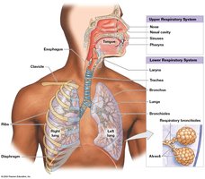
Functional Divisions
Conducting Portion: Nasal cavity to larger bronchioles (Most of the upper and lower respiratory system)
Respiratory Portion: Smallest bronchioles and alveoli ( air filled pockets in lungs for site of gas exchange)
Respiratory Mucosa and Defense
The respiratory mucosa lines the conducting portion and consists of respiratory epithelium and lamina propria. It plays a key role in trapping and removing debris and pathogens.
Respiratory Epithelium: Varies by location; includes pseudostratified ciliated columnar epithelium, stratified squamous epithelium, and cuboidal epithelium.
Lamina Propria: Contains mucous glands in upper regions and smooth muscle in lower regions.
Mucociliary Escalator: Cilia move mucus and trapped particles toward the pharynx for swallowing.
Alveolar Macrophages: Engulf small particles in alveoli.
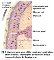
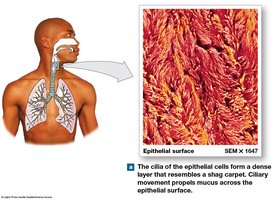
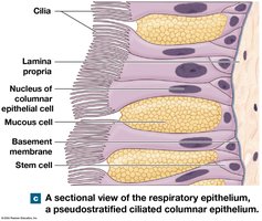
Respiratory Defense System = a series of filtration mechanisms that remove debris and pathogens from the inhaled air.
Mucus: Traps particles and pathogens.
Cilia: Sweep mucus toward the pharynx.
Macrophages: Remove debris in alveoli.
Clinical Conditions: Overload ( (A lot of airborne particles not allowed to enter into the respiratory system like lungs causes an overload of respiratory defenses (mucus plugs and inflammation(smoking)), tuberculosis (bacterial infection in the lungs = fever, coughing, chest pain), cystic fibrosis (congenital defect affecting mucus producing cells = thick sticky mucus, blocking respiratory passageways and frequent infections).
Upper Respiratory System
Nose and Nasal Cavity
The nose and nasal cavity are the entry points for air and play roles in filtration, humidification, and olfaction.
Nasal Vestibule: Entry space; nasal hairs trap large particles.
Nasal Septum: Divides cavity; formed by vomer, ethmoid, and cartilage.
Olfactory Region: Superior area lined by olfactory epithelium for smell.
Conchae and Meatuses: Create turbulent airflow for warming, humidifying, and olfactory stimuli to reach olfactory receptors, and trap particles. (Conchae = superior, middle and inferior conchae) (Air flows between the conchae through the superior, middle and inferior nasal meatuses)
–Hard palate – forms the floor of nasal cavity
▪Separates the nasal and oral cavities
▪Consists of the maxillae and palatine bones
–Soft palate – extends posterior to hard palate and divides the superior nasopharynx from the rest of the pharynx
–Choanae – posterior opening of the nasal cavity into the pharynx
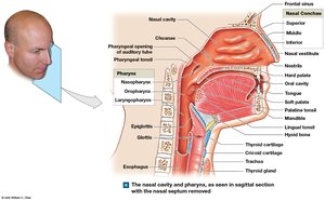
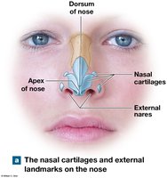
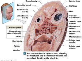
Palate and Pharynx (a chamber shared by the digestive and respiratory systems)
Pharynx: Shared chamber for respiratory and digestive systems; divided into nasopharynx (superior portion), oropharynx (posterior to oral cavity), and laryngopharynx (inferior portion between hyoid bone and entrance of larynx and esophagus lined with stratified squamous epithelium to resist abrasion).
Lower Respiratory System
Larynx Structure and Function
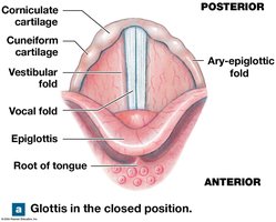
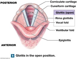
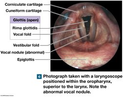
The larynx = cartilaginous structure involved in breathing and sound production. Air passes from the pharynx into the larynx through the glottis (slit-like opening between vocal cords).
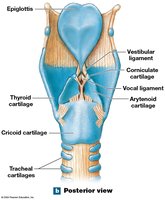
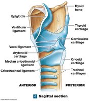
Thyroid Cartilage (hyaline cartilage): Forms anterior/lateral walls; laryngeal prominence (Adam's apple).
Cricoid Cartilage (hyaline cartilage): Forms posterior portion .
Epiglottis (elastic cartilage): Covers glottis during swallowing. Larynx elevates and epiglottis folds back over glottis to block food and liquids from entering in the respiratory tract.
Arytenoid, Corniculate, Cuneiform Cartilages: Involved in glottis movement and sound production.
Cartilages = support and structure.
Ligaments = connect cartilages. (Connect and support the cartilages, help hold the vocal folds in place, assist with opening and closing the glottis.)
Vocal folds = produce sound.
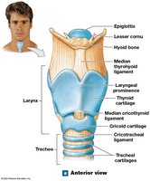
Ligaments and Vocal Folds
Vestibular Ligaments: Protect vocal folds.
Vocal Ligaments: Elastic, involved in sound production.
Trachea and Bronchial Tree
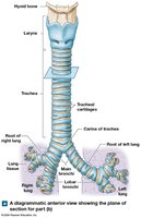
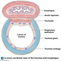
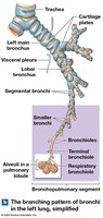
The trachea is a flexible tube supported by C-shaped cartilages. It branches into the right and left main bronchi, which further divide into lobar and segmental bronchi.
Tracheal Cartilages: Keep airway open; allow flexibility during swallowing.
Bronchial Tree: Progressive branching; less cartilage, more smooth muscle in smaller bronchi.
Bronchioles: Smallest branches; dominated by smooth muscle.
Bronchodilation: Increases airway diameter (sympathetic).
Bronchoconstriction: Decreases diameter (parasympathetic/allergic reactions).
Gas Exchange Structures
Alveolar Anatomy
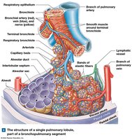
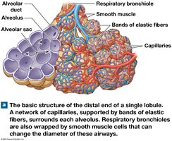
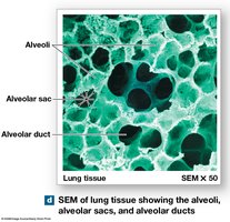
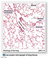
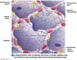
Alveoli are the primary sites of gas exchange. Each terminal bronchiole forms several respiratory bronchioles, which connect to alveoli via alveolar ducts and sacs.
Pneumocytes Type I: Simple squamous cells for gas diffusion.
Pneumocytes Type II: Produce surfactant to reduce surface tension.
Alveolar Macrophages: Patrol alveolar surfaces.
Blood-Air Barrier
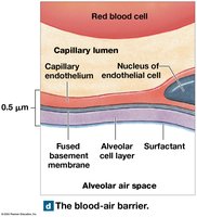
Layers: Alveolar cell layer, capillary endothelial layer, fused basement membrane.
Efficiency: Short diffusion distance, lipid-soluble gases.
Clinical: Pneumonia can compromise the barrier.
The Lungs
Superficial Anatomy
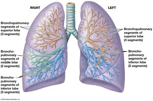
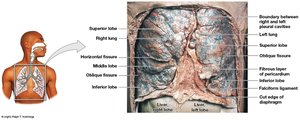
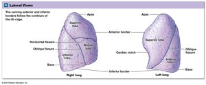
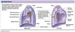
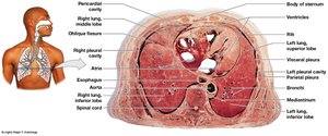
The lungs are located in pleural cavities and divided into lobes by fissures. The right lung has three lobes; the left lung has two and a cardiac notch.
Hilum: Entry/exit for vessels, nerves, bronchi.
Root: Dense connective tissue anchoring structures.
Pulmonary Lobules and Blood Supply
Pulmonary Lobules: Small segments divided by trabeculae and interlobular septa.
Pulmonary Circuit: Deoxygenated blood arrives via arteries; oxygenated blood returns via veins.
Systemic Circuit: Supplies conducting portions.
Pulmonary Embolism: Blockage of pulmonary vessels can cause tissue collapse or heart failure.
Pleural Cavities
Parietal Pleura: Lines thoracic wall.
Visceral Pleura: Covers lungs.
Pleural Fluid: Lubricates cavity.
Pleurisy: Pain/inflammation due to reduced lubrication.
Respiration Processes
External vs. Internal Respiration
Respiration includes external (exchange between environment and tissues) and internal (cellular absorption/release) processes.
External Respiration: Pulmonary ventilation, gas diffusion, transport in blood.
Internal Respiration: Oxygen absorption and carbon dioxide release by cells.
Pulmonary Ventilation
Physical Principles and Muscle Actions
Pulmonary ventilation is the movement of air in and out of the lungs, governed by pressure gradients and Boyle's law.
Boyle's Law: (Pressure inversely proportional to volume)
Air Movement: Flows from high to low pressure.
Muscles: Diaphragm and external intercostals for inhalation; internal intercostals and abdominal muscles for forceful exhalation.
Pressure Changes
Atmospheric Pressure: 1 atm = 760 mm Hg
Intrapulmonary Pressure: Small changes during relaxed breathing; larger during forceful breathing.
Intrapleural Pressure: Always lower than atmospheric; assists venous return.
Respiratory Rates and Volumes
Respiratory Rate: 12-18 breaths/min (adult)
Tidal Volume (TV): 500 ml
Minute Volume:
Alveolar Ventilation: Adjusted for dead space
Pulmonary Volumes and Capacities
Inspiratory Reserve Volume (IRV): Extra inhaled air
Expiratory Reserve Volume (ERV): Extra exhaled air
Residual Volume: Air remaining after maximal exhalation
Vital Capacity: Maximum air moved in/out
Total Lung Capacity: Total lung volume
Gas Exchange
Physical Principles
Partial Pressure: Each gas contributes to total pressure (Dalton's law)
Henry's Law: Gas solubility in liquid proportional to partial pressure
Solubility: CO2 highly soluble; O2 less; N2 limited
External Respiration
Oxygen: Diffuses from alveoli to blood
Carbon Dioxide: Diffuses from blood to alveoli
Internal Respiration
Oxygen: Diffuses from blood to tissues
Carbon Dioxide: Diffuses from tissues to blood
Gas Transport
Hemoglobin and Oxygen Transport
Oxygen: 1.5% dissolved in plasma; remainder binds to hemoglobin (Hb)
Oxyhemoglobin: Hb with bound O2
Hemoglobin Saturation: % of heme units with O2
Factors: Partial pressure, pH (Bohr effect), temperature, BPG levels
Fetal Hemoglobin
Higher affinity for O2 than adult Hb
Carbon Dioxide Transport
Bicarbonate Ions: 70% converted in RBCs
Carbaminohemoglobin: 23% bound to Hb
Dissolved in Plasma: 7%
Control of Respiration
Regulation and Reflexes
Local Regulation: Tissue CO2 and O2 levels affect diffusion and blood flow.
Neural Control: Respiratory centers in medulla and pons regulate rate and depth.
Voluntary Control: Cerebral cortex can override involuntary centers.
Respiratory Reflexes
Chemoreceptors: Respond to CO2, O2, pH
Baroreceptors: Respond to blood pressure changes
Stretch Receptors: Respond to lung volume changes
Protective Reflexes: Sneezing, coughing, laryngeal spasm
Age-Related Changes in Respiration
Newborns and Aging
Newborns: Lungs inflate at birth; foramen ovale and ductus arteriosus close.
Aging: Elastic tissue deteriorates, compliance decreases, vital capacity lowers, chest flexibility reduces, emphysema risk increases.
Integration with Other Systems
Respiratory and Cardiovascular Coordination
Homeostasis: Maintained by coordination with cardiovascular system.
Reflexes: Chemoreceptor and baroreceptor reflexes affect both systems.
Additional info: This study guide covers the main anatomical and physiological principles of the respiratory system, including clinical correlations and regulatory mechanisms, suitable for college-level Anatomy & Physiology students.
