 Indietro
Chapter 5: The Integumentary System – Anatomy & Physiology Study Notes
Guida di studio - Note intelligenti
 Appunti personalizzati basati sui tuoi materiali, ampliati con definizioni chiave, esempi e contesto.
The Integumentary System
Overview and Functions
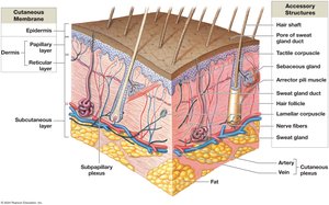
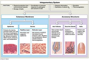
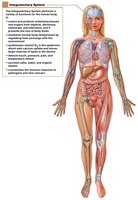
The integumentary system is the largest organ system in the human body, comprising about 16% of body weight and covering 1.5–2 square meters. It consists of the cutaneous membrane (skin), which includes the epidermis and dermis, as well as accessory structures such as hair, nails, and exocrine glands. The system also contains blood vessels and sensory receptors, and is separated from deeper tissues by the subcutaneous layer (hypodermis).
Protection: Shields underlying tissues from impact, abrasion, fluid loss, and chemical attack.
Excretion: Removes salts, water, and organic wastes via glands.
Temperature Regulation: Maintains body temperature through insulation and evaporative cooling.
Melanin Production: Protects against UV radiation.
Keratin Production: Provides toughness and water resistance.
Vitamin D Synthesis: Essential for calcium metabolism.
Lipid Storage: Stores lipids in dermis and subcutaneous layer.
Sensory Detection: Detects touch, pressure, pain, vibration, and temperature.
Immune Response: Coordinates defense against pathogens and cancers.
Epidermis
Structure and Function
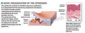
The epidermis is the superficial layer of the skin, composed of stratified squamous epithelium. It is avascular, relying on diffusion from capillaries in the dermis for nutrients and oxygen. The main cell type is the keratinocyte, which produces keratin, a tough, fibrous protein.
Thin skin: Covers most of the body; has four layers.
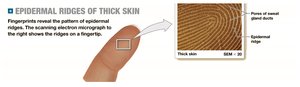
Thick skin: Covers palms and soles; has five layers.
Layers of the Epidermis
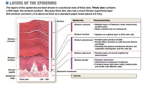
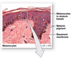
Stratum basale: Deepest layer; contains stem cells (basal cells), tactile (Merkel) cells, and melanocytes. Attached to basement membrane by hemidesmosomes. Forms epidermal ridges (fingerprints).
Stratum spinosum: 8–10 layers of keratinocytes bound by desmosomes; contains dendritic (Langerhans) cells for immune defense.
Stratum granulosum: 3–5 layers; cells stop dividing, fill with keratin and keratohyalin, become dehydrated and die.
Stratum lucidum: Only in thick skin; layer of dead keratinocytes.
Stratum corneum: 15–30 layers; keratinized cells, water-resistant but not waterproof; cells shed after 2 weeks.
Specialized Cells and Growth
Tactile (Merkel) cells: Sensory receptors for touch, found in hairless skin.
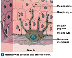
Melanocytes: Synthesize melanin pigment, distributed in stratum basale.
Epidermal Growth Factor (EGF): Promotes division of basal cells, keratin production, epidermal development, and repair.
Dermis
Structure and Function
The dermis is the deeper layer of skin, anchoring accessory structures and containing blood vessels and nerves. It is divided into two layers:
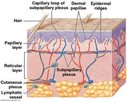
Papillary layer: Areolar tissue; contains capillaries, lymphatic vessels, and sensory neurons; forms dermal papillae.
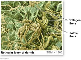
Reticular layer: Dense irregular connective tissue; rich in collagen and elastic fibers; provides strength and flexibility.
Dermal Strength, Elasticity, and Blood Supply
Collagen fibers: Strong, limit flexibility.
Elastic fibers: Allow stretching and recoil.
Skin turgor: Strength and flexibility due to water content.
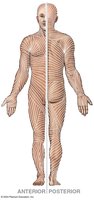
Tension lines: Parallel bundles of fibers; cuts parallel heal better.
Cutaneous plexus: Deep network of vessels.
Subpapillary plexus: Network in papillary layer.
Innervation
Tactile (Meissner) corpuscles: Detect light touch.
Lamellar (Pacinian) corpuscles: Detect deep pressure and vibration.
Subcutaneous Layer (Hypodermis)
Structure and Function
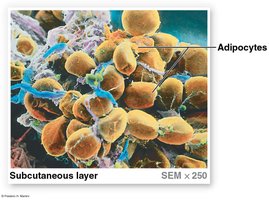
The subcutaneous layer stabilizes the skin and connects it to underlying tissues. It is not part of the skin, but consists mainly of adipose tissue for energy storage, insulation, and padding. It contains large arteries and veins and is a site for subcutaneous injections.
Skin Color
Factors Affecting Skin Color
Carotene: Orange-yellow pigment from vegetables; accumulates in skin and can be converted to vitamin A.
Melanin: Red-yellow (pheomelanin) or brown-black (eumelanin) pigment produced by melanocytes; protects against UV radiation.
Dermal circulation: Blood flow and oxygenation affect skin color; cyanosis indicates low oxygen.
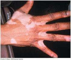
Disease-related changes: Jaundice, pituitary tumors, Addison’s disease, and vitiligo can alter skin color.
Vitamin D3 Synthesis
Sunlight and Vitamin D Production
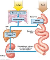
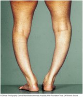
Vitamin D3 (cholecalciferol) is produced by epidermal cells in response to UV radiation. The liver and kidneys convert it to calcitriol, which is essential for calcium and phosphate absorption in the small intestine. Deficiency can cause rickets, resulting in abnormal bone development.
Hair
Structure and Function
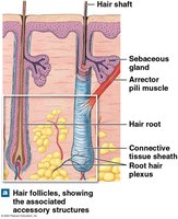
Hair is a nonliving accessory structure produced in hair follicles, originating in the dermis and projecting through the epidermis. It covers most of the body except certain areas.
Functions: Protection, cushioning, insulation, guarding openings, and sensory reception.
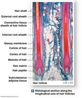
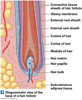
Hair follicles: Originate deep in dermis, surrounded by connective tissue sheath and sensory nerves (root hair plexus).
Arrector pili muscle: Causes "goose bumps" by contracting.
Hair Structure and Growth
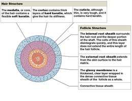
Hair root: Anchors hair in skin.
Hair shaft: Visible portion.
Medulla: Central core.
Cortex: Intermediate layer.
Cuticle: Surface layer.
Internal root sheath: Surrounds hair root.
External root sheath: Extends from skin surface to hair matrix.
Glassy membrane: Clear layer wrapped in connective tissue sheath.
Hair production: Occurs at hair bulb; cells keratinize and die as they move upward.
Hair growth cycle: Includes growth, club hair formation, and shedding.
Types of hair: Vellus (soft, fine) and terminal (heavy, pigmented).
Hair color: Determined by melanocyte activity, genes, environment, and age.
Exocrine Glands of the Skin
Types and Functions
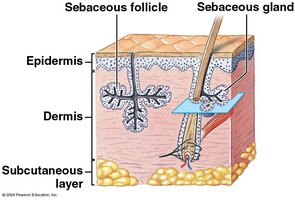
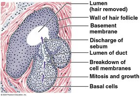
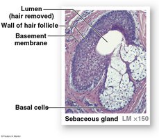
Sebaceous glands: Produce sebum, lubricate and protect hair, inhibit bacteria.
Sebaceous follicles: Discharge sebum directly onto skin surface.
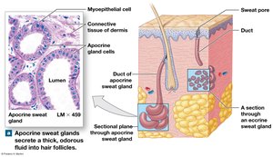
Apocrine sweat glands: Secrete into hair follicles; produce sticky, odorous secretions.
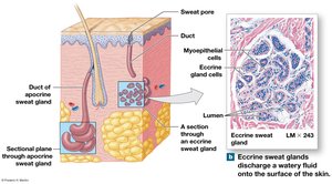
Eccrine (merocrine) sweat glands: Secrete directly onto skin; regulate temperature, excrete water/electrolytes, protect from hazards.
Mammary glands: Produce milk.
Ceruminous glands: Produce earwax (cerumen).
Control of Glandular Secretions
Autonomic nervous system: Controls sebaceous and apocrine glands.
Eccrine glands: Controlled more precisely; main function is thermoregulation.
Nails
Structure and Formation
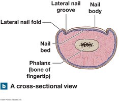
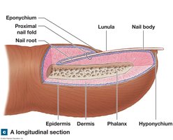
Nails are protective structures made of dead, keratinized epidermal cells. They cover the tips of fingers and toes.
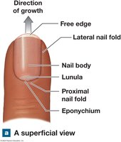
Nail body: Visible portion.
Nail bed: Underlying tissue.
Hyponychium: Thickened stratum corneum beneath free edge.
Nail root: Site of nail production.
Eponychium (cuticle): Stratum corneum over nail root.
Lunula: Pale crescent near root.
Injury Repair
Phases of Skin Injury Repair
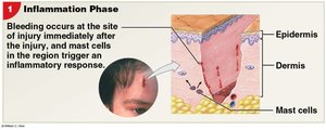
Inflammatory phase: Bleeding, swelling, pain; mast cells trigger inflammation.
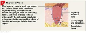
Migration phase: Scab forms; macrophages clean debris; cells migrate to repair epidermis and dermis.
Proliferation phase: Scab disintegrates; fibroblasts produce collagen meshwork; epidermal cells migrate.
Scarring phase: Scar tissue forms; less flexible, more fibers, fewer blood vessels.
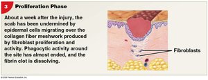
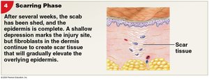
Scar Formation
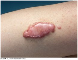
Keloid: Excessive scar tissue, thick and raised, covered by shiny epidermis.
Effects of Aging on the Integumentary System
Changes with Age
Epidermis thins: Connections between dermis and epidermis weaken.
Dendritic cells decrease: Increased infection risk.
Vitamin D production declines.
Melanocyte activity declines: Increased risk of burns and cancers.
Glandular activity declines: Loss of lubrication and cooling ability.
Blood supply to dermis reduced: Decreased thermoregulation.
Hair follicle function declines: Hair loss, graying.
Elastic fiber network shrinks: Sagging and wrinkling.
Repair rate slows.
Clinical Notes
Skin Cancer
Basal cell carcinoma: Originates in stratum basale; most common; does not metastasize.
Squamous cell carcinoma: Affects squamous cells; does not metastasize.
Malignant melanoma: Aggressive cancer of melanocytes; metastasis common.
Burns and Grafts
First-degree: Partial-thickness, damages only epidermis.
Second-degree: Partial-thickness, damages epidermis and some dermis.
Third-degree: Full-thickness, destroys epidermis, dermis, and subcutaneous layer; requires grafts.
Rule of nines: Used to estimate burn surface area.
Complications: Fluid/electrolyte loss, thermoregulation issues, increased infection risk.
