 Indietro
Chapter 5: The Integumentary System – Anatomy & Physiology Study Guide
Guida di studio - Note intelligenti
 Appunti personalizzati basati sui tuoi materiali, ampliati con definizioni chiave, esempi e contesto.
The Integumentary System
Introduction to the Integumentary System
The integumentary system is the body's largest organ system, consisting of the skin and its derivatives (hair, nails, and glands). It serves as a protective barrier, regulates temperature, and provides sensory information.
Skin: Composed of two main layers—the epidermis and dermis—with a subcutaneous layer (hypodermis) beneath.
Appendages: Includes hair, nails, sweat glands, and sebaceous glands.
Functions: Protection, sensation, thermoregulation, metabolic functions, blood reservoir, and excretion.
Structure of the Skin
Layers and Components of Skin
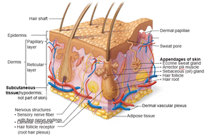
The skin is organized into distinct layers, each with specialized structures and functions. The epidermis is the outermost layer, followed by the dermis, and then the hypodermis (subcutaneous tissue).
Epidermis: Stratified squamous epithelium; avascular.
Dermis: Connective tissue; contains blood vessels, nerves, and skin appendages.
Hypodermis: Not part of skin; consists mainly of adipose tissue.
Appendages: Eccrine sweat gland, arrector pili muscle, sebaceous gland, hair follicle, hair root.
Nervous Structures: Sensory nerve fibers, lamellar corpuscles, hair follicle receptors.
Epidermis: Cells and Layers
Layers of the Epidermis
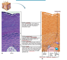
The epidermis consists of several layers, each with unique cell types and functions. Cell differentiation occurs as cells move from the basal layer to the surface.
Stratum Basale: Deepest layer; contains actively dividing keratinocytes and melanocytes.
Stratum Spinosum: Several layers of keratinocytes; contains dendritic cells.
Stratum Granulosum: Cells contain keratohyalin granules; begin to die.
Stratum Lucidum: Present only in thick skin (palms, soles); clear layer.
Stratum Corneum: Outermost layer; dead, flattened keratinocytes provide protection.
Dermis and Skin Color
Papillary and Reticular Layers of Dermis
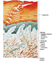
The dermis is divided into two layers: the papillary layer (loose connective tissue) and the reticular layer (dense irregular connective tissue). These layers provide structural support and house important skin features.
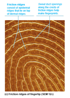
Papillary Layer: Contains dermal papillae, capillaries, and sensory receptors.
Reticular Layer: Contains collagen fibers, elastic fibers, and cleavage lines.
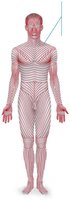
Cleavage Lines: Natural lines formed by collagen fiber orientation; important for surgical incisions.
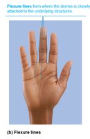
Flexure Lines: Form where the dermis is tightly attached to underlying structures (e.g., palms).
Dermal Modifications and Clinical Imbalances
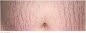
Dermal modifications can result in characteristic skin markings, such as stretch marks (striae) and blisters.
Striae: Silvery white scars from dermal tears due to extreme stretching.
Blisters: Fluid-filled pockets separating epidermal and dermal layers due to acute trauma.
Skin Color: Key Pigments
Skin color is determined by three main pigments: melanin, carotene, and hemoglobin. Excessive sun exposure can damage skin and increase risk of cancer.
Melanin: Produced by melanocytes; responsible for brown-black color.
Carotene: Yellow-orange pigment from diet; accumulates in stratum corneum.
Hemoglobin: Red pigment in blood; gives pinkish hue to fair skin.
Clinical Imbalances: Excessive UV exposure can cause DNA damage, folic acid destruction, and photosensitivity.
Hair: Structure and Functions
Hair Composition and Follicle Structure
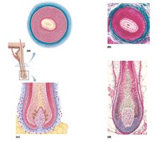
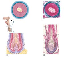
Hair is composed of dead, keratinized cells and grows from follicles embedded in the dermis. The follicle structure is complex, with several layers and associated cells.
Hair Shaft: Visible part above skin surface.
Hair Root: Part within the follicle.
Follicle Wall: Includes peripheral connective tissue sheath, glassy membrane, epithelial root sheath.
Hair Matrix: Actively dividing cells responsible for hair growth.
Melanocytes: Provide pigment to hair.
Cutaneous Glands
Sweat Glands and Sebaceous (Oil) Glands
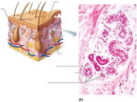
Cutaneous glands include sweat glands (eccrine and apocrine) and sebaceous glands. These glands play roles in thermoregulation, lubrication, and protection.
Eccrine Sweat Glands: Produce watery sweat for cooling; found throughout body.
Apocrine Sweat Glands: Found in axillary and genital areas; produce thicker secretion.
Sebaceous Glands: Secrete sebum (oil) into hair follicles; lubricates skin and hair.
Clinical Imbalances: Acne, whiteheads, blackheads, and seborrhea (cradle cap) are common disorders.
Functions of the Skin
Protection, Regulation, and Sensation
The skin provides multiple protective barriers, regulates body temperature, and enables cutaneous sensations.
Protection: Physical, chemical, and biological barriers.
Body Temperature Regulation: Sweat production and blood flow adjustments.
Cutaneous Sensations: Touch, pressure, pain, and temperature detection via specialized receptors.
Metabolic Functions: Synthesis of vitamin D, chemical conversions.
Blood Reservoir: Stores blood in dermal vessels.
Excretion: Eliminates waste products via sweat.
Skin Cancer and Burns
Skin Cancer
Skin cancer is a common disorder, often caused by excessive UV exposure. Types include basal cell carcinoma, squamous cell carcinoma, and melanoma.
Basal Cell Carcinoma: Most common, least malignant.
Squamous Cell Carcinoma: Can metastasize if not treated.
Melanoma: Most dangerous; arises from melanocytes.
Burns: Classification and Severity
Burns are classified by depth and extent of tissue damage. The Rule of Nines is used to estimate burn severity and affected body surface area.
First-Degree Burn: Only epidermis; redness and pain.
Second-Degree Burn: Epidermis and upper dermis; blisters.
Third-Degree Burn: Entire thickness of skin; risk of fluid loss and infection.
Rule of Nines: Divides body into regions, each representing 9% (or multiples) of total body surface area.
Burn Degree | Layers Affected | Symptoms |
|---|---|---|
First | Epidermis | Redness, pain |
Second | Epidermis & upper dermis | Blisters, pain |
Third | Entire skin | No pain (nerve endings destroyed), risk of infection |
Body Region | % of Total Surface Area |
|---|---|
Head & Neck | 9% |
Upper Limbs | 18% |
Trunk | 36% |
Lower Limbs | 36% |
Perineum | 1% |
Key Takeaways
The integumentary system is essential for protection, regulation, and sensation.
Skin structure includes the epidermis, dermis, and hypodermis, with specialized appendages.
Clinical imbalances can affect skin health and function.
Understanding skin cancer and burns is critical for clinical practice.
