 Indietro
Chapter 6: The Skeletal System – Anatomy & Physiology Study Guide
Guida di studio - Note intelligenti
 Appunti personalizzati basati sui tuoi materiali, ampliati con definizioni chiave, esempi e contesto.
The Skeletal System
Overview and Components
The skeletal system is a complex framework that provides structural support, protection, and movement for the human body. It consists of bones, cartilages, joints, ligaments, and connective tissues that stabilize or connect bones.
Bones: The main structural elements.
Cartilages: Provide flexibility and cushioning.
Joints: Allow movement between bones.
Ligaments: Connect bones and stabilize joints.
Connective tissue: Supports and connects skeletal elements.
Functions of the Skeletal System
The skeletal system performs several essential functions:
Support: Provides structural support for the entire body; bones serve as attachment points for soft tissues and organs.
Storage of minerals and lipids: Bones store calcium and phosphorus ions; yellow bone marrow stores lipids for energy.
Blood cell production: Red and white blood cells, as well as platelets, are produced in red bone marrow.
Protection: Skeletal structures surround and protect vital organs (e.g., skull protects the brain, ribs protect heart and lungs).
Leverage: Bones act as levers, changing the size and direction of force generated by muscles, resulting in movement.
Bone Tissue and Structure
Bone Tissue Characteristics
Bone, or osseous tissue, is a supporting connective tissue containing specialized cells (osteocytes) embedded in a matrix of calcium salts and collagen fibers.
Osteocytes: Mature bone cells responsible for maintaining bone structure.
Matrix: Composed of calcium phosphate (Ca3(PO4)2) and collagen fibers.
Classification of Bones by Shape
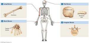
Bones are classified based on their shape:
Long bones: Longer than they are wide (e.g., humerus).
Short bones: About as wide as they are long (e.g., carpal bones).
Flat bones: Thin and broad (e.g., parietal bone).
Irregular bones: Complex shapes (e.g., vertebrae).
Features of a Long Bone
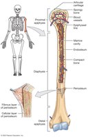
Long bones have distinct structural features:
Diaphysis: Central shaft surrounding the marrow cavity.
Epiphyses: Expanded ends covered with articular cartilage, articulating with adjacent bones.
Periosteum: Outer covering with fibrous and cellular layers.
Endosteum: Inner lining covering spongy bone and marrow cavity.
Types of Bone Tissue
Compact bone: Dense and solid, forms the wall of the diaphysis.
Spongy bone: Network of trabeculae, fills epiphyses and lines marrow cavity.
Microscopic Features of Bone
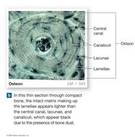
Bone cells (osteocytes) are located in lacunae, between lamellae (calcified matrix). Canaliculi are small channels connecting lacunae to blood vessels for nutrient and waste exchange.
Histology of Compact Bone
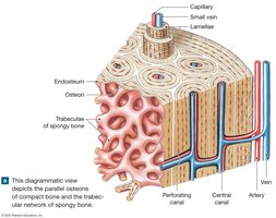
Osteon (Haversian system): Basic functional unit; osteocytes arranged in concentric lamellae around a central canal.
Central canal: Contains blood vessels.
Perforating canals: Link blood vessels of central canal with periosteum and marrow cavity.
Structural Features of Spongy Bone
Trabeculae: Rods or plates forming the network.
Red bone marrow: Found in spaces between trabeculae; site of blood cell production.
Types of Bone Cells
Osteoblasts: Produce new bone (ossification).
Osteocytes: Mature cells maintaining bone structure.
Osteoclasts: Break down bone matrix (resorption).
Bone Formation and Growth
Ossification Processes
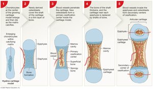
Bone formation begins in the embryo as cartilage is replaced by bone. Two main processes:
Intramembranous ossification: Bone develops within sheets of fibrous connective tissue (e.g., flat bones of skull).
Endochondral ossification: Bone replaces hyaline cartilage models (most bones).
Appositional Growth
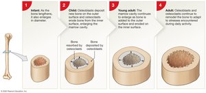
Growth in bone diameter occurs as periosteum cells develop into osteoblasts, producing matrix on the outer surface, while osteoclasts erode the inner surface, enlarging the marrow cavity.
Requirements for Bone Growth
Minerals: Calcium and phosphate.
Vitamin D3: Essential for calcium metabolism.
Vitamin A and C: Support osteoblasts.
Hormones: Growth hormone, thyroid hormone, sex hormones, and calcium-balancing hormones (PTH and calcitonin).
Bone Remodeling
Bone remodeling is a continuous process involving osteocytes, osteoclasts, and osteoblasts. It allows bones to adapt to new stresses and maintain strength.
Heavily stressed bones: Become thicker and stronger.
Inactivity: Leads to thinner, more brittle bones.
Calcium Regulation
Calcium is the most abundant mineral in the body, with 99% stored in bones. Regulation is critical for physiological processes.
Parathyroid hormone (PTH) and calcitriol: Raise blood calcium levels.
Calcitonin: Lowers blood calcium levels.
Skeletal Divisions and Bone Markings
Axial and Appendicular Skeletons
The human skeleton is divided into two main parts:
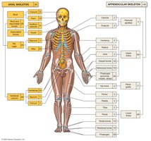
Axial skeleton: Skull, rib cage, and vertebral column (80 bones).
Appendicular skeleton: Pectoral and pelvic girdles, arms, and legs (126 bones).
Bone Markings
Bone markings are surface features that serve as landmarks for muscle attachment, articulation, and passage of blood vessels and nerves.
Elevations/processes: Attachment points for tendons and ligaments.
Depressions/grooves/openings: Passageways for blood vessels and nerves.
Axial Skeleton: Skull, Vertebral Column, and Thoracic Cage
Skull Bones
The skull consists of cranial and facial bones, protecting the brain and supporting sensory organs.
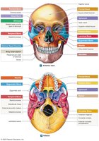
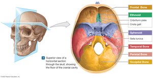
Frontal bone: Forms forehead and roof of orbits.
Parietal bones: Form roof and superior walls of cranium.
Occipital bone: Forms posterior and inferior cranium; contains foramen magnum.
Temporal bones: Form sides of cranium; house auditory ossicles.
Sphenoid bone: Floor of cranium; contains sella turcica.
Ethmoid bone: Forms cranial floor, medial orbit, and nasal cavity.
Facial Bones
Maxillary bones: Largest facial bones; form palate and orbit.
Palatine bones: Form posterior palate.
Zygomatic bones: Form cheekbones.
Nasal and lacrimal bones: Form bridge of nose and medial orbit.
Vomer: Forms part of nasal septum.
Mandible: Lower jaw; only movable facial bone.
Paranasal Sinuses and Nasal Complex
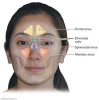
Paranasal sinuses are air-filled cavities that lighten skull weight and produce mucus. The nasal septum separates right and left nasal cavities.
The Hyoid Bone
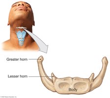
The hyoid bone is U-shaped, not attached to other bones, and serves as a base for muscles of the larynx, tongue, and throat.
Infant Skull and Fontanelles
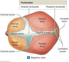
Fontanelles are areas of fibrous connective tissue in infant skulls, allowing for growth and easier delivery. They ossify by age four.
Vertebral Column
The vertebral column consists of 26 bones: 24 vertebrae, the sacrum, and the coccyx. It provides support and protects the spinal cord.
Cervical region: 7 vertebrae (C1–C7).
Thoracic region: 12 vertebrae (T1–T12).
Lumbar region: 5 vertebrae (L1–L5).
Sacral region: 5 fused vertebrae (sacrum).
Coccygeal region: 3–5 fused vertebrae (coccyx).
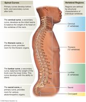
Spinal Curvature
Primary curves: Present at birth (thoracic and sacral).
Secondary curves: Develop after birth (cervical and lumbar).
Abnormal curvatures: Kyphosis, lordosis, scoliosis.
Vertebral Bone Markings
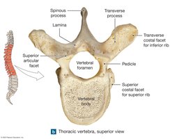
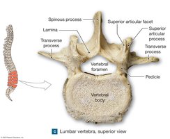
Vertebral body: Weight-bearing portion.
Vertebral canal: Formed by vertebral foramen.
Transverse and spinous processes: Muscle attachment sites.
Articular processes: Form joints between vertebrae.
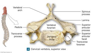
Distinctive Features of Vertebrae
Cervical: Small body, large foramen, transverse foramina.
Atlas (C1): Supports skull, allows nodding.
Axis (C2): Dens allows rotation.
Thoracic: Heart-shaped body, costal facets for ribs.
Lumbar: Largest, thickest body, stumpy spinous process.
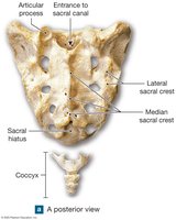
Sacrum: Triangular, fused vertebrae, pelvic attachment.
Coccyx: Fused vertebrae, muscle attachment.
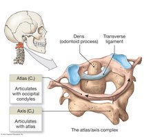
Thoracic Cage
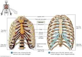
The thoracic cage consists of thoracic vertebrae, ribs, and sternum, protecting vital organs and supporting respiration.
Ribs: 12 pairs; true ribs (1–7), false ribs (8–12), floating ribs (11–12).
Sternum: Manubrium, body, xiphoid process.
Appendicular Skeleton: Pectoral and Pelvic Girdles, Limbs
Pectoral Girdle
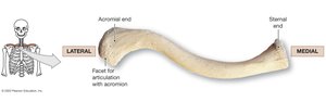
Clavicle: S-shaped bone connecting sternum and scapula.
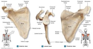
Scapula: Triangular bone with glenoid cavity, coracoid process, acromion, and scapular spine.
Upper Limb
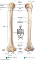
Humerus: Arm bone; articulates with scapula and forearm.
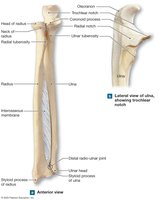
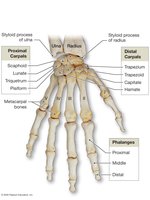
Radius and ulna: Forearm bones; radius lateral, ulna medial.
Carpals, metacarpals, phalanges: Bones of wrist, hand, and fingers.
Pelvic Girdle
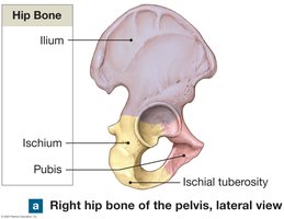
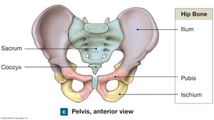
Coxal bones: Fusion of ilium, ischium, and pubis.
Pelvis: Includes hip bones, sacrum, and coccyx; stabilized by ligaments.
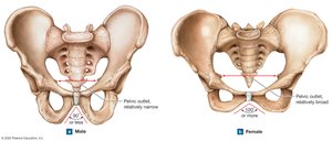
Sex differences: Female pelvis is broader, with larger outlet and pubic angle.
Lower Limb
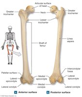
Femur: Thigh bone; longest and heaviest.
Patella: Kneecap; sesamoid bone.
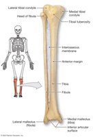
Tibia and fibula: Leg bones; tibia is medial and weight-bearing, fibula is lateral and slender.
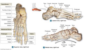
Tarsals, metatarsals, phalanges: Bones of ankle, foot, and toes.
Joints and Articulations
Classification of Joints
Joints are classified by structure and function:
Structural: Fibrous, cartilaginous, synovial.
Functional: Synarthrosis (immovable), amphiarthrosis (slightly movable), diarthrosis (freely movable).
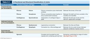
Functional Category | Structural Category and Type | Description | Example |
|---|---|---|---|
Synarthrosis (No Movement) | Fibrous - Suture Cartilaginous - Synchondrosis | Fibrous connections; plate interlocked surfaces Cartilaginous connection | Between skull bones Between first rib and sternum |
Amphiarthrosis (Little Movement) | Fibrous - Syndesmosis Cartilaginous - Symphysis | Ligamentous connection Connection by fibrocartilage pad | Between tibia and fibula Between pubic bones |
Diarthrosis (Free Movement) | Synovial | Complex joint surrounded by joint capsule and containing synovial fluid | Shoulder, knee, hip |
Synovial Joints
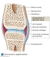
Articular cartilage: Covers bone ends.
Joint capsule: Surrounds joint.
Synovial membrane: Lines capsule, produces synovial fluid.
Synovial fluid: Lubricates, nourishes cartilage, reduces friction.
Types of Synovial Joint Movement
Plane (gliding): Surfaces slide past each other.
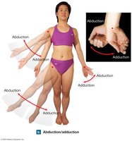
Angular: Flexion, extension, hyperextension, abduction, adduction, circumduction.
Rotational: Rotation, pronation, supination.
Special: Inversion, eversion, dorsiflexion, plantar flexion, opposition, reposition, elevation, depression, lateral flexion.
Types of Synovial Joints
Plane (gliding): Slight movement (carpals).
Hinge: Angular movement in one plane (elbow, knee).
Saddle: Angular movement in two planes (thumb).
Pivot: Rotation only (atlas-axis).
Condylar (ellipsoidal): Angular movement in two planes (wrist).
Ball-and-socket: Movement in all planes (shoulder, hip).
Representative Joints
Shoulder: Ball-and-socket, greatest range of motion, rotator cuff muscles.
Elbow: Hinge joint, stable, strong ligaments.
Hip: Ball-and-socket, extremely stable, strong ligaments and muscles.
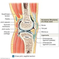
Knee: Hinge and plane joints, menisci, ligaments, fat pads, bursae.
Functional Relationships with Other Body Systems
The skeletal system interacts with muscular, cardiovascular, lymphatic, endocrine, digestive, and urinary systems for movement, mineral balance, and physiological regulation.
Summary Table: Key Bone and Joint Features
Bone Type | Example | Function |
|---|---|---|
Long | Humerus, femur | Leverage, movement |
Short | Carpals, tarsals | Stability, support |
Flat | Parietal, sternum | Protection |
Irregular | Vertebrae | Complex functions |
Joint Type | Example | Movement |
|---|---|---|
Hinge | Elbow, knee | Flexion/extension |
Ball-and-socket | Shoulder, hip | All planes |
Pivot | Atlas-axis | Rotation |
Plane | Carpals | Gliding |
Additional info: Tables and images have been recreated and expanded for clarity. All key terms and concepts are explained for self-contained study.
