 Indietro
Chapter 7: The Nervous System – Structured Study Notes
Guida di studio - Note intelligenti
 Appunti personalizzati basati sui tuoi materiali, ampliati con definizioni chiave, esempi e contesto.
The Nervous System
Overview and Functions
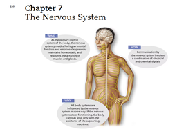
The nervous system is a complex network responsible for regulating and coordinating body activities. It enables sensory input, integration of information, and motor output, which together maintain homeostasis and control voluntary and involuntary actions.
Sensory Input: Sensory receptors detect changes (stimuli) inside and outside the body.
Integration: The nervous system processes and interprets sensory input, deciding if action is needed.
Motor Output: A response activates muscles or glands.
Feedback Loop: These functions mirror a feedback loop, where receptors send input to the brain (control center), which processes and determines the output.
Organization of the Nervous System
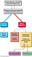
The nervous system is organized structurally and functionally. Structural classification divides it into the central and peripheral nervous systems, while functional classification distinguishes sensory and motor divisions.
Central Nervous System (CNS): Consists of the brain and spinal cord; serves as the integration and command center.
Peripheral Nervous System (PNS): Composed of nerves extending from the CNS; connects sensory organs, muscles, and glands.
Sensory (Afferent) Division: Carries information to the CNS from sensory receptors.
Motor (Efferent) Division: Carries impulses from the CNS to effector organs.
Structural Classification
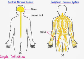
Structural classification is based on the anatomical components of the nervous system.
CNS: Brain and spinal cord; interprets sensory information and issues instructions.
PNS: Nerves (cranial and spinal) that serve as communication lines between the CNS and the rest of the body.
Functional Classification
Functional classification is based on the direction and type of information flow.
Sensory (Afferent) Division: Somatic fibers carry information from skin, muscles, and joints; visceral fibers from internal organs.
Motor (Efferent) Division: Somatic nervous system controls voluntary skeletal muscles; autonomic nervous system controls involuntary actions (smooth/cardiac muscle, glands), further divided into sympathetic and parasympathetic systems.
Nervous Tissue: Structure and Function
Cell Types in Nervous Tissue
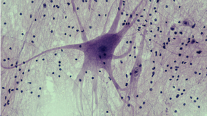
Nervous tissue consists of two main cell types: neurons and neuroglia (supporting cells).
Neurons: Specialized cells that transmit electrical impulses.
Neuroglia: Support, insulate, and protect neurons; unable to conduct impulses and retain the ability to divide.
Types of Neuroglia
Neuroglia are classified based on their location and function.
CNS Neuroglia:
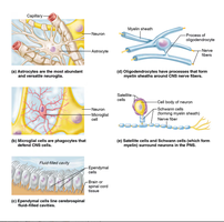
Astrocytes: Anchor neurons to capillaries, regulate exchanges, and control the chemical environment.
Microglia: Phagocytes that monitor neuron health and dispose of debris.
Ependymal Cells: Line brain and spinal cord cavities; cilia circulate cerebrospinal fluid.
Oligodendrocytes: Produce myelin sheaths around CNS axons.
PNS Neuroglia:
Schwann Cells: Form myelin sheaths in the PNS.
Satellite Cells: Protect and cushion neuron cell bodies.
Neurons: Structure and Properties
Neuron Anatomy
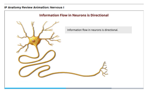
Neurons are specialized for transmitting messages. They consist of a cell body and processes (dendrites and axons).
Cell Body: Contains nucleus and metabolic machinery.
Dendrites: Receive impulses and conduct them toward the cell body.
Axon: Conducts impulses away from the cell body; ends in axon terminals containing neurotransmitters.
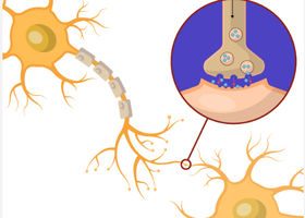
Synaptic Cleft: Gap between axon terminals and the next neuron.
Synapse: Functional junction for impulse transmission.
Myelin Sheaths
Myelin is a white, fatty substance that insulates axons and speeds up impulse transmission.
Schwann Cells: Form myelin in the PNS.
Oligodendrocytes: Form myelin in the CNS.
Neuron Terminology
Nuclei: Clusters of cell bodies in the CNS.
Ganglia: Collections of cell bodies in the PNS.
Tracts: Bundles of nerve fibers in the CNS.
Nerves: Bundles of nerve fibers in the PNS.
White Matter: Myelinated fibers.
Gray Matter: Unmyelinated fibers and cell bodies.
Functional Classification of Neurons
Neurons are classified by the direction of information flow.
Sensory (Afferent) Neurons: Carry impulses from receptors to the CNS.
Motor (Efferent) Neurons: Carry impulses from the CNS to muscles/glands.
Interneurons: Connect sensory and motor neurons within the CNS.
Structural Classification of Neurons
Multipolar: Many extensions; most common, includes motor and interneurons.
Bipolar: One axon and one dendrite; found in special sense organs.
Unipolar: Single process; sensory neurons in PNS ganglia.
Functional Properties of Neurons
Irritability: Ability to respond to a stimulus and convert it to a nerve impulse.
Conductivity: Ability to transmit the impulse to other neurons, muscles, or glands.
Electrical Activity of Neurons
Resting Membrane Potential
The plasma membrane of a resting neuron is polarized, with fewer positive ions inside than outside.
Major Positive Ion Inside: Potassium (K+)
Major Positive Ion Outside: Sodium (Na+)
Equation:
Action Potential Initiation and Generation
A stimulus changes membrane permeability, allowing sodium ions to enter and causing depolarization.
Depolarization: Membrane potential changes from negative to positive (e.g., −60 mV to +40 mV).
Action Potential: If depolarization is sufficient, an action potential is generated and propagated.
Equation:
Propagation and Repolarization
Propagation: Action potential travels along the axon; myelinated fibers conduct faster.
Repolarization: Restores the inside to a negative charge and outside to positive.
Synaptic Transmission
Steps of Synaptic Transmission
Neurons communicate at synapses through a series of steps involving electrical and chemical signals.
Action potential reaches axon terminal, opening calcium channels.
Calcium causes vesicles with neurotransmitters to fuse with the membrane.
Neurotransmitters are released into the synaptic cleft.
Neurotransmitters bind to receptors on the next neuron.
If enough neurotransmitter is released, a graded potential is generated, leading to an action potential.
Neurotransmitter is quickly removed by reuptake or enzymatic activity.
Reflexes and Reflex Arcs
Reflexes
Reflexes are rapid, predictable, and involuntary responses to stimuli, occurring over neural pathways called reflex arcs.
Somatic Reflexes: Stimulate skeletal muscles (e.g., withdrawal from pain).
Autonomic Reflexes: Regulate smooth muscle, heart, glands (e.g., blood pressure regulation).
Elements of a Reflex Arc
Sensory Receptor: Reacts to stimulus.
Sensory Neuron: Carries message to CNS.
Integration Center: Processes information.
Motor Neuron: Carries message to effector.
Effector Organ: Muscle or gland stimulated.
Types of Reflex Arcs
Two-Neuron Reflex Arc: Simplest type; example is the patellar reflex.
Three-Neuron Reflex Arc: Includes receptor, sensory neuron, interneuron, motor neuron, and effector; example is the flexor (withdrawal) reflex.
Summary Table: Structural and Functional Classification of Nervous System
Classification | Components | Function |
|---|---|---|
Central Nervous System (CNS) | Brain, Spinal Cord | Integration, command center |
Peripheral Nervous System (PNS) | Cranial and Spinal Nerves | Communication lines |
Sensory (Afferent) Division | Sensory fibers | Carry information to CNS |
Motor (Efferent) Division | Motor fibers | Carry impulses from CNS to effectors |
Somatic Nervous System | Skeletal muscles | Voluntary control |
Autonomic Nervous System | Cardiac/smooth muscle, glands | Involuntary control |
