 Indietro
Comprehensive Study Notes: The Reproductive System (Anatomy & Physiology)
Guida di studio - Note intelligenti
 Appunti personalizzati basati sui tuoi materiali, ampliati con definizioni chiave, esempi e contesto.
The Reproductive System
Overview and Objectives
The reproductive system is essential for the continuation of species, involving both male and female anatomical structures, gamete production, hormonal regulation, and developmental processes. Key objectives include understanding the anatomy of both systems, meiosis, hormonal regulation, development, puberty, and menopause.
Anatomy: Study of male and female reproductive organs
Meiosis: Process of gamete formation
Hormonal Regulation: Control of reproductive functions
Development: Formation and maturation of reproductive systems
Puberty & Menopause: Life stage changes in reproductive function
Primary Sex Organs and Accessory Structures
The primary sex organs, or gonads, are the testes in males and ovaries in females. Gonads produce gametes (sperm and ova) and secrete sex hormones. Accessory organs include ducts, glands, and external genitalia.
Sex Hormones: Androgens (e.g., testosterone), estrogens, and progesterone
Functions: Development and function of reproductive organs, sexual behavior, and drives
Male Reproductive System
Anatomy of the Male Reproductive System
The male reproductive system consists of internal and external structures, including the testes, scrotum, penis, and accessory glands.
Testes: Produce sperm and androgens
Scrotum: External sac that houses the testes, regulates temperature for optimal sperm production
Penis: Organ of copulation, contains erectile tissue (corpus spongiosum and corpora cavernosa)
Accessory Glands: Seminal vesicles, prostate gland, bulbourethral glands
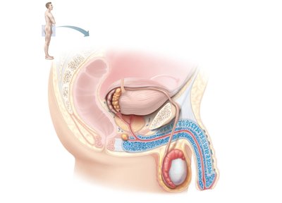
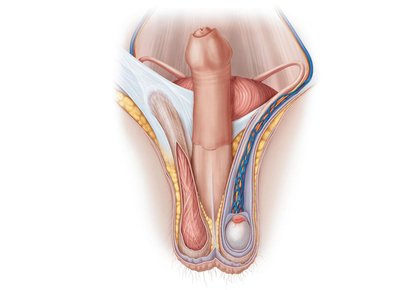
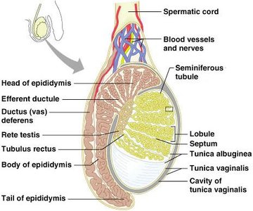
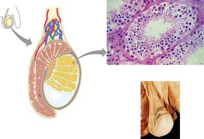
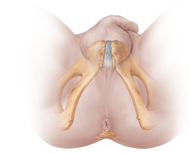
Testes and Scrotum
The testes are covered by the tunica vaginalis and tunica albuginea. Seminiferous tubules within the testes produce sperm, while interstitial (Leydig) cells produce androgens. The scrotum maintains a temperature lower than core body temperature, essential for spermatogenesis.
Spermatic Cord: Encloses vas deferens, nerves, blood vessels, and lymphatics
Pampiniform Plexus: Venous network that helps regulate testicular temperature

Penis
The penis consists of a root, shaft, and glans. The prepuce (foreskin) covers the glans. Erectile tissue enables erection during sexual arousal.
Corpus Spongiosum: Surrounds the urethra
Corpora Cavernosa: Paired columns of erectile tissue

Epididymis, Ductus Deferens, and Urethra
Epididymis: Site of sperm maturation and storage
Ductus (Vas) Deferens: Transports sperm from epididymis to ejaculatory duct
Urethra: Conveys urine and semen; divided into prostatic, membranous, and spongy regions
Accessory Glands
Seminal Vesicles: Secrete 60% of semen volume, provide nutrients
Prostate Gland: Secretes milky, slightly acidic fluid, activates sperm
Bulbourethral Glands: Produce mucus to neutralize acidic urine
Semen
Semen is a mixture of sperm and glandular secretions, providing a transport medium, nutrients, and protection for sperm.
Hormone Relaxin: Enhances sperm motility
Alkalinity: Neutralizes acidic environment
Prostaglandins: Facilitate sperm movement through female tract
Cell Division: Mitosis and Meiosis
Comparison of Mitosis and Meiosis
Mitosis and meiosis are processes of cell division. Mitosis produces identical diploid cells for growth and repair, while meiosis produces haploid gametes for reproduction, introducing genetic variability.
Mitosis: One division, produces two identical diploid cells
Meiosis: Two divisions, produces four genetically unique haploid cells
Synapsis and Crossover: Occur in meiosis, increasing genetic diversity



Spermatogenesis
Process of Spermatogenesis
Spermatogenesis is the sequence of events that produces sperm in the seminiferous tubules. It involves mitosis, meiosis, and spermiogenesis.
Spermatogonia: Stem cells undergo mitosis
Primary Spermatocytes: Undergo meiosis I
Secondary Spermatocytes: Undergo meiosis II
Spermatids: Resulting haploid cells, transform into spermatozoa
Spermiogenesis
Spermiogenesis is the transformation of spermatids into functional sperm, involving the development of the acrosome, flagellum, and condensation of nuclear material.
Acrosome: Contains enzymes for egg penetration
Flagellum: Provides motility
Midpiece: Contains mitochondria for energy
Sertoli (Sustentacular) Cells
Sertoli cells support and nourish developing sperm, form the blood-testis barrier, and regulate spermatogenesis.
Blood-Testis Barrier: Protects sperm from immune response
Compartmentalization: Basal and adluminal compartments
Hormonal Regulation of Male Reproduction
Hypothalamic-Pituitary-Gonadal Axis
The hypothalamic-pituitary-gonadal axis regulates testicular function through hormones.
GnRH: Stimulates release of FSH and LH from anterior pituitary
FSH: Stimulates Sertoli cells and spermatogenesis
LH: Stimulates Leydig cells to produce testosterone
Inhibin: Provides negative feedback
Testosterone and Male Secondary Sex Characteristics
Testosterone is synthesized from cholesterol and is responsible for male secondary sex characteristics, libido, and maintenance of reproductive organs.
Pubic, axillary, and facial hair
Deepening of voice
Increased muscle and bone mass
Female Reproductive System
Anatomy of the Female Reproductive System
The female reproductive system includes the ovaries, uterine tubes, uterus, vagina, and external genitalia (vulva).
Ovaries: Primary sex organs, produce ova and hormones
Uterine Tubes: Transport ova to uterus
Uterus: Site of implantation and fetal development
Vagina: Organ of copulation and birth canal
External Genitalia: Vulva, including labia majora, labia minora, clitoris
Ovarian Follicles and Oogenesis
Oogenesis is the production of female gametes by meiosis. Follicles develop through several stages: primordial, primary, secondary, and vesicular (Graafian).
Oogonia: Stem cells multiply by mitosis
Primary Oocytes: Begin meiosis, stall in prophase I
Secondary Oocytes: Complete meiosis I, arrest in metaphase II
Ovulation: Release of secondary oocyte
Uterus and Endometrium
The uterus is supported by ligaments and consists of three layers: perimetrium, myometrium, and endometrium. The endometrium undergoes cyclic changes in response to ovarian hormones.
Stratum Functionalis: Shed during menstruation
Stratum Basalis: Regenerates functionalis
Vascular Supply: Uterine, arcuate, and radial arteries
External Genitalia and Mammary Glands
Vulva: Includes labia majora, labia minora, clitoris, and vestibule
Mammary Glands: Modified sweat glands, produce milk
Ovarian and Uterine Cycles
Ovarian Cycle
The ovarian cycle consists of the follicular phase (follicle growth), ovulation, and luteal phase (corpus luteum activity).
Follicular Phase: Days 1–14, follicle growth
Ovulation: Day 14, release of secondary oocyte
Luteal Phase: Days 14–28, corpus luteum secretes hormones
Hormonal Regulation
GnRH: Stimulates FSH and LH release
FSH and LH: Stimulate follicle growth and hormone production
Estrogen: Inhibits FSH/LH at low levels, positive feedback at high levels
Progesterone: Maintains endometrium
Uterine Cycle
The uterine cycle consists of three phases:
Menstrual Phase: Shedding of endometrial functional layer
Proliferative Phase: Rebuilding of endometrium
Secretory Phase: Preparation for embryo implantation
Developmental Aspects
Genetic Sex Determination
Genetic sex is determined by X and Y chromosomes. The SRY gene on the Y chromosome initiates testes development.
Gonadal Ridges: Form in the 5th week
Male Structures: Develop in the 7th week
Female Structures: Develop in the 8th week
Development of Internal and External Genitalia
Male: Genital tubercle forms penis, labioscrotal swellings form scrotum
Female: Genital tubercle forms clitoris, labioscrotal swellings form labia majora
Descent of Gonads
Testes descend into the scrotum before birth, guided by the gubernaculum. Ovaries descend but are stopped by the broad ligament.
Puberty and Menopause
Puberty: Reproductive organs mature, secondary sex characteristics appear
Menopause: Cessation of ovulation and menses, decline in estrogen
Sexually Transmitted Diseases (STDs)
Bacterial Infections
Gonorrhea: Painful urination, pus discharge
Syphilis: Can be congenital, treated with penicillin
Chlamydia: Most common STD, can cause sterility
Viral Infections
Genital Warts: Caused by HPV, increases cancer risk
Genital Herpes: Latent periods, can cause fetal malformations
Summary Table: Comparison of Mitosis and Meiosis
Feature | Mitosis | Meiosis |
|---|---|---|
Number of Divisions | One | Two |
Synapsis of Homologous Chromosomes | No | Yes |
Daughter Cells | Two, diploid, identical | Four, haploid, genetically unique |
Role | Growth, repair | Gamete production |
Key Equations
Chromosomal Number: (diploid), (haploid)
Meiosis:
Additional info:
Some images were omitted due to lack of direct anatomical or physiological relevance.
Tables and diagrams were recreated for clarity and completeness.
