 Indietro
Digestive System: Structure and Function Study Guide
Guida di studio - Note intelligenti
 Appunti personalizzati basati sui tuoi materiali, ampliati con definizioni chiave, esempi e contesto.
Digestive System Overview
General Structure and Function
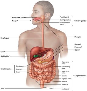
The digestive system, also known as the alimentary canal, gastrointestinal (GI) tract, or gut, is responsible for the breakdown and absorption of nutrients, as well as the elimination of waste. It includes accessory organs such as the liver and pancreas.
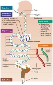
Ingestion: Intake of food into the mouth.
Propulsion: Movement of food through the GI tract via peristalsis.
Mechanical Breakdown: Physical breakdown of food by chewing and churning.
Segmentation: Mixing of food in the small intestine.
Digestion: Chemical breakdown of food by enzymes.
Absorption: Uptake of nutrients into the body.
Defecation: Elimination of indigestible substances as feces.
Anatomy of the Alimentary Canal
Peritoneum and Mesenteries
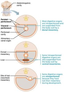
The peritoneum is a serous membrane lining the abdominal cavity and covering the digestive organs.
Visceral Peritoneum: Covers the external surfaces of digestive organs.
Parietal Peritoneum: Lines the inside of the abdominal cavity.
Peritoneal Cavity: Space between the two peritoneal layers, filled with peritoneal fluid.
Mesenteries: Double layers of peritoneum that suspend organs and provide routes for blood vessels, nerves, and lymphatics.
Histology of the Alimentary Canal
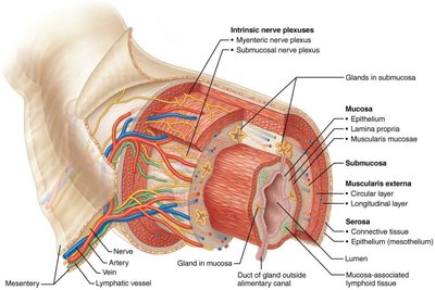
The wall of the alimentary canal consists of four layers, each with distinct functions.
Mucosa: Innermost layer; secretes enzymes and mucus, absorbs nutrients, and protects against infection.
Submucosa: Areolar connective tissue containing blood vessels, nerves, and lymphatics.
Muscularis Externa: Smooth muscle responsible for peristalsis and segmentation.
Serosa: Outermost layer; provides protection and structural support.
Functional Anatomy of the Digestive System
Mouth and Associated Structures
The oral cavity is lined with mucus and contains glands, teeth, and the tongue, which work together to form a bolus for swallowing.
Lips and Cheeks: Keep food in the mouth.
Palate: Hard (anterior) and soft (posterior) palates separate the oral and nasal cavities.
Tongue: Muscular organ that mixes food and aids in swallowing.
Salivary Glands: Parotid, sublingual, and submandibular glands produce saliva containing amylase and lipase.
Teeth: Incisors, canines, premolars, and molars perform cutting, tearing, and grinding.
Pharynx and Esophagus
Pharynx: Oropharynx and laryngopharynx transport food to the esophagus.
Esophagus: Muscular tube that carries food to the stomach via peristalsis; lined with stratified squamous epithelium for protection.
Stomach
Structure and Function
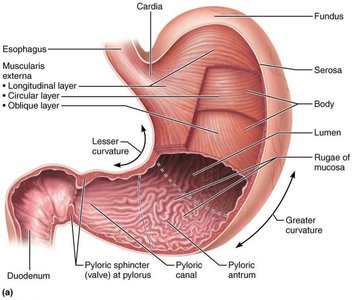
The stomach serves as a storage site, mechanically breaks down food, and initiates protein digestion.
Capacity: 50 ml (empty) to 4 L (full).
Sphincters: Cardiac sphincter (entry), pyloric sphincter (exit).
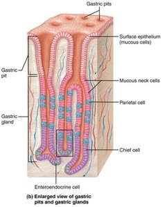
Gland Cells: Mucous neck cells (acidic mucus), parietal cells (HCl and intrinsic factor), chief cells (pepsinogen and lipases), enteroendocrine cells (hormones).
Mucosal Barrier and Digestive Processes
The stomach is protected from its own acidic secretions by a mucosal barrier.
Bicarbonate-rich mucus: Neutralizes acid.
Tight junctions: Prevent leakage of gastric juice.
Cell turnover: Constant replacement of epithelial cells.
Ulcers: Caused by Helicobacter pylori destroying mucus.
Phases of Gastric Secretion
Cephalic Phase: Triggered by sight, smell, or thought of food.
Gastric Phase: Initiated when food enters the stomach.
Intestinal Phase: Begins as food enters the duodenum.
Alkaline Tide: Increase in blood pH due to HCO3- ions entering the blood.
Gastric Motility and Emptying
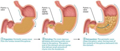
The stomach mixes food into chyme and regulates its release into the duodenum.
Receptive Relaxation: Allows stomach to expand.
Mixing: Smooth muscle churns food.
Pylorus: Squirts small volumes of chyme into the duodenum.
Emptying Time: 4 hours for carb-rich meals, 6+ hours for fatty/solid meals.
Liver, Gallbladder, and Pancreas
Liver
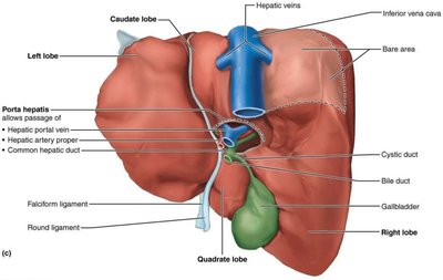
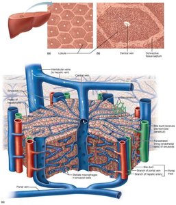
The liver is a large gland with four lobes, responsible for producing bile, processing nutrients, storing vitamins, and detoxifying blood.
Blood Supply: Hepatic artery and hepatic portal vein supply blood; hepatic vein drains blood.
Bile Ducts: Common hepatic duct joins cystic duct from gallbladder to form bile duct.
Microscopic Anatomy of the Liver
Hepatocytes: Arranged in lobules; perform metabolic and detoxification functions.
Hepatic Macrophages: Clean blood.
Bile: Alkaline fluid containing bile salts and phospholipids; emulsifies lipids.
Homeostatic Disorders: Hepatitis (inflammation), cirrhosis (chronic inflammation), gallstones (imbalance of bile salts and cholesterol).
Gallbladder
Function: Stores and releases bile.
Gallstones: Formed from too few bile salts or too much cholesterol.
Pancreas
Exocrine Function: Acinar cells produce pancreatic juice, rich in digestive enzymes.
Enzymes: Proteases (trypsinogen, carboxypeptidase, chymotrypsin), amylase (starch), lipases (fats), nucleases (nucleic acids).
Activation: Trypsinogen is converted to trypsin by enteropeptidase in the duodenum.
Small Intestine
Structure and Function
The small intestine is the major digestive organ, where most absorption occurs.
Duodenum: 25 cm; receives secretions from liver and pancreas.
Jejunum: 2.5 m; primary site of absorption.
Ileum: 3.6 m; ends at ileocecal sphincter.
Microscopic Anatomy
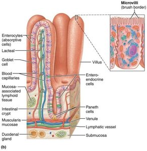
Villi and Microvilli: Increase surface area for absorption.
Intestinal Crypts: Produce secretions.
Enterocytes: Absorptive cells.
Goblet Cells: Produce mucus.
Paneth Cells: Secrete antimicrobial enzymes.
Stem Cells: Generate new epithelial cells.
MALT: Includes Peyer's patches for immune defense.
Duodenal Glands: Produce alkaline mucus.
Digestive Processes in the Small Intestine
Bile and Enzymes: Imported from liver and pancreas.
Segmentation: Mixes chyme.
Peristalsis: Moves chyme along.
Absorption: Most nutrients and water absorbed; lipids absorbed by lacteals and enter lymphatic system.
Large Intestine
Structure and Function
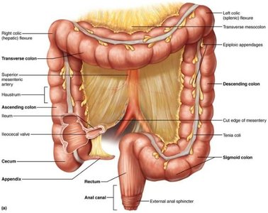
The large intestine absorbs water and some nutrients, forms and stores feces, and eliminates waste.
Sections: Cecum (with appendix), colon (ascending, transverse, descending, sigmoid), rectum, anal canal, anus.
Appendix: Houses beneficial bacteria and lymphoid tissue.
Bacterial Microbiota: Over 1,000 species; commensal, mutualistic, and pathogenic types; synthesize vitamins B and K; produce gases.
Digestive Processes of the Large Intestine
Conversion: Chyme converted to feces over 12-24 hours.
Absorption: Water, vitamins, and electrolytes absorbed.
Defecation Reflex: Triggered by rectal wall stretching; internal anal sphincter (involuntary), external anal sphincter (voluntary).
Summary Table: Layers of the Alimentary Canal
Layer | Main Function | Key Components |
|---|---|---|
Mucosa | Secretion, absorption, protection | Epithelium, lamina propria, muscularis mucosae |
Submucosa | Support, supply | Areolar connective tissue, blood vessels, nerves, lymphatics |
Muscularis Externa | Motility (peristalsis, segmentation) | Circular and longitudinal smooth muscle layers |
Serosa | Protection | Connective tissue, mesothelium |
Key Equations
Alkaline Tide: Increase in blood pH due to bicarbonate ion release:
Additional info:
Digestive system processes are tightly regulated by neural and hormonal mechanisms to ensure efficient breakdown and absorption of nutrients.
Accessory organs (liver, pancreas, gallbladder) play crucial roles in digestion by producing and delivering enzymes and bile.
