 Indietro
Endocrine System: Structure, Function, and Regulation
Guida di studio - Note intelligenti
 Appunti personalizzati basati sui tuoi materiali, ampliati con definizioni chiave, esempi e contesto.
Endocrine System Overview
Introduction to the Endocrine System
The endocrine system is a network of glands that produce and secrete hormones to regulate various bodily functions, including metabolism, growth, reproduction, and homeostasis. Hormones are chemical messengers that travel through the bloodstream to target organs and tissues.
Key Glands: Pituitary, thyroid, parathyroid, adrenal, pancreas, gonads, pineal, thymus.
Hormones: Regulate processes such as metabolism, stress response, blood glucose, and calcium balance.
Regulation: Feedback mechanisms (positive and negative) control hormone release.
Hormones of the Posterior Pituitary
Oxytocin
Oxytocin is a peptide hormone released by the posterior pituitary. It is a strong stimulant of uterine contraction and is regulated by a positive feedback mechanism.
Functions: Stimulates uterine contractions during labor; triggers milk ejection (letdown reflex) in lactating women; involved in sexual arousal and satisfaction.
Clinical Use: Synthetic and natural oxytocic drugs are used to induce or hasten labor.
Antidiuretic Hormone (ADH)
ADH helps prevent dehydration by reducing urine formation. It acts on the kidneys to promote water reabsorption.
Mechanism: Stimulates aquaporin installation in collecting ducts, allowing water reabsorption and concentrating urine.
Regulation: Osmoreceptors monitor blood solute concentration; high solutes trigger ADH release, low solutes inhibit it.
Inhibition: Alcohol inhibits ADH, causing increased urine output.
Thyroid Gland
Anatomy and Histology
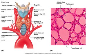
The thyroid gland is the largest endocrine gland, located in the anterior neck. It consists of two lateral lobes connected by the isthmus and is composed of follicles that produce thyroglobulin.
Follicles: Produce thyroglobulin, a precursor to thyroid hormones.
Colloid: Mixture of thyroglobulin and iodine, fills the follicle lumen.
Parafollicular Cells: Produce calcitonin.
Thyroid Hormones: T3 and T4
Thyroid hormones are major metabolic regulators.
T4 (Thyroxine): Two tyrosine molecules plus four iodine atoms; 90% of thyroid hormone secreted.
T3 (Triiodothyronine): Two tyrosines plus three iodine atoms; 10% of thyroid hormone secreted.
Effects of Thyroid Hormone
Metabolic Rate: Increases glucose oxidation, metabolic rate, and heat production (thermogenesis).
Other Roles: Maintains blood pressure, regulates tissue growth, develops skeletal and nervous systems, and supports reproductive capabilities.
Synthesis of Thyroid Hormone
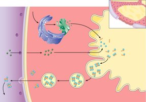
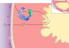
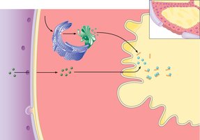
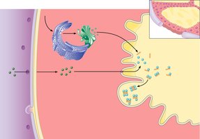
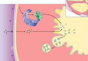
The synthesis of thyroid hormone involves several steps:
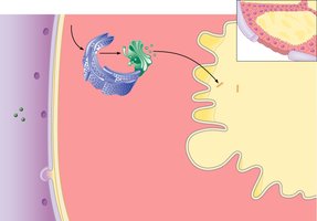
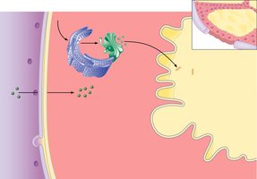
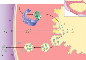
Thyroglobulin is synthesized and discharged into the follicle lumen.
Iodide (I–) is actively transported into the cell, oxidized to iodine (I2), and released into the lumen.
Iodine attaches to tyrosine, forming monoiodotyrosine (MIT) and diiodotyrosine (DIT).
Iodinated tyrosines link to form T3 and T4.
Colloid is endocytosed and combined with a lysosome, where T3 and T4 are cleaved and diffuse into the bloodstream.
Cellular Effects of Thyroid Hormone
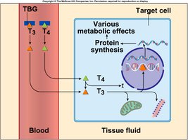
Binding: TH binds to receptors on mitochondria (increases aerobic respiration), ribosomes, and chromatin (increases protein synthesis).
Na+-K+ ATPase: TH stimulates production of sodium-potassium pump, generating heat.
T4 Conversion: T4 is converted to T3 in peripheral tissues.
Endemic Goiter
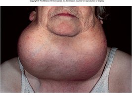
Endemic goiter is caused by insufficient iodine in the diet, leading to thyroid gland hypertrophy due to lack of negative feedback.
Pathophysiology: Without iodine, TH cannot be synthesized, resulting in increased TRH and TSH secretion and gland enlargement.
Prevention: Iodized salt is used to prevent goiter.
Transport and Regulation of Thyroid Hormone
Transport: T4 and T3 bind to thyroxine-binding globulins (TBGs) produced by the liver.
Activity: T3 is ten times more active than T4; peripheral tissues convert T4 to T3.
Regulation: Negative feedback; hypothalamic TRH can override feedback.
Calcitonin
Calcitonin is a peptide hormone produced by parafollicular cells.
Function: Lowers blood calcium levels, especially in children.
Mechanism: Inhibits osteoclast activity and stimulates calcium uptake into bone matrix.
Regulation: Humoral negative feedback based on blood calcium concentration.
Parathyroid Glands
Anatomy and Histology
Parathyroid glands are tiny glands embedded in the posterior aspect of the thyroid. Chief cells secrete parathyroid hormone (PTH). 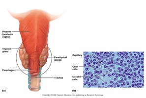
Effects of Parathyroid Hormone (PTH)
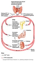
PTH regulates calcium balance in the blood.
Stimulates osteoclasts: Digests bone matrix, releasing calcium.
Enhances kidney reabsorption: Increases Ca2+ reabsorption and phosphate excretion.
Increases intestinal absorption: Promotes Ca2+ absorption by intestinal mucosa.
Regulation: Rising Ca2+ inhibits PTH release (humoral mechanism).
Adrenal (Suprarenal) Glands
Anatomy and Histology
Adrenal glands are paired, pyramid-shaped organs atop the kidneys. They consist of the adrenal cortex (glandular tissue) and adrenal medulla (neural tissue). 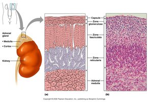
Adrenal Cortex: Corticosteroids
The adrenal cortex synthesizes and releases steroid hormones called corticosteroids.
Zona glomerulosa: Mineralocorticoids (chiefly aldosterone).
Zona fasciculata: Glucocorticoids (chiefly cortisol).
Zona reticularis: Gonadocorticoids (chiefly androgens).
Mineralocorticoids (Aldosterone)
Function: Regulates electrolytes in extracellular fluids; maintains Na+ balance by reducing sodium excretion and stimulating reabsorption in kidneys.
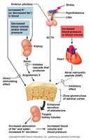
Regulation: Stimulated by rising K+, low Na+, decreased blood volume/pressure.
Mechanisms of Aldosterone Secretion
Renin-angiotensin mechanism
Plasma concentration of sodium and potassium
ACTH (during stress)
Atrial natriuretic peptide (ANP) inhibits aldosterone
Glucocorticoids (Cortisol)
Function: Helps resist stress by keeping blood sugar levels constant and maintaining blood volume.
Effects: Provokes gluconeogenesis, increases blood glucose, fatty acids, and amino acids.
Excess: Depresses cartilage/bone formation, inhibits inflammation, depresses immune system, and affects cardiovascular, neural, and GI function.
Gonadocorticoids (Sex Hormones)
Androgens: Contribute to puberty, secondary sex characteristics, and sex drive in females.
Estrogens: Small amounts produced, especially after menopause.
Adrenal Medulla
Chromaffin cells: Secrete epinephrine and norepinephrine.
Effects: Increase blood glucose, constrict blood vessels, increase heart rate, divert blood to brain, heart, and skeletal muscle.
Epinephrine: More potent for heart and metabolism.
Norepinephrine: More influential on vasoconstriction and blood pressure.
Stress Response (General Adaptation Syndrome)
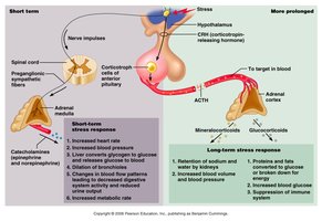
Alarm Reaction: Norepinephrine and epinephrine prepare body for fight-or-flight; depletes glycogen.
Stage of Resistance: Cortisol breaks down fat and protein for gluconeogenesis; immune system inhibited.
Stage of Exhaustion: Fat depleted, muscle wasting, rapid decline, and death.
Pancreas
Anatomy and Function
The pancreas is a triangular gland with both exocrine and endocrine functions.
Acinar cells: Produce digestive enzymes (exocrine).
Pancreatic islets: Produce hormones (endocrine).
Alpha cells: Produce glucagon.
Beta cells: Produce insulin.
Glucagon
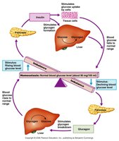
Glucagon is a potent hyperglycemic agent.
Target: Liver.
Actions: Promotes glycogenolysis, gluconeogenesis, and release of glucose into blood.
Insulin
Insulin lowers blood glucose levels and enhances glucose transport into cells.
Actions: Catalyzes glucose oxidation for ATP, polymerizes glucose to glycogen, converts glucose to fat.
Receptor: Tyrosine kinase enzyme.
Regulation of Blood Glucose Levels
Glucagon: Raises blood glucose.
Insulin: Lowers blood glucose.
Diabetes Mellitus (DM)
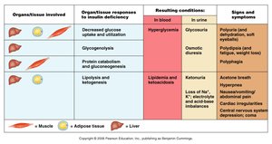
Diabetes mellitus results from hyposecretion or hypoactivity of insulin.
Type I: Juvenile onset, insulin deficiency.
Type II: Adult onset, insulin resistance.
Cardinal Signs: Polyuria (excess urine), polydipsia (excess thirst), polyphagia (excess hunger).
Hyperinsulinism: Excessive insulin secretion causes hypoglycemia.
Gonads
Female Gonads (Ovaries)
Ovaries produce estrogens and progesterone, responsible for maturation of reproductive organs, secondary sexual characteristics, breast development, and cyclic changes in uterine mucosa.
Male Gonads (Testes)
Testes produce testosterone, which initiates maturation of male reproductive organs, secondary sexual characteristics, sex drive, sperm production, and maintenance of sex organs.
Pineal Gland
Function
The pineal gland produces melatonin, which regulates day/night cycles (circadian rhythm) and physiological processes with rhythmic variations.
Thymus
Function
The thymus produces thymopoietins and thymosins, essential for T lymphocyte development and immune function.
Other Hormone-Producing Structures
Heart: Produces atrial natriuretic peptide (ANP) to reduce blood pressure, volume, and sodium concentration.
Gastrointestinal tract: Enteroendocrine cells release local digestive hormones.
Placenta: Releases hormones influencing pregnancy.
Kidneys: Secrete erythropoietin for red blood cell production.
Skin: Produces cholecalciferol (vitamin D precursor).
Adipose tissue: Releases leptin for satiety and energy expenditure.
