Indietro
Fundamentals of the Nervous System: Structure and Function
Guida di studio - Note intelligenti
 Appunti personalizzati basati sui tuoi materiali, ampliati con definizioni chiave, esempi e contesto.
Nervous System Basics
Divisions of the Nervous System
The nervous system is divided into two main parts: the Central Nervous System (CNS) and the Peripheral Nervous System (PNS). Each division has distinct structures and functions essential for communication and control throughout the body.
CNS: Consists of the brain and spinal cord. Responsible for integration and interpretation of sensory input and dictating motor responses.
PNS: Composed of cranial and spinal nerves and their branches. Transports sensory input toward the CNS and motor output away from the CNS.
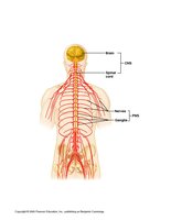
Functional Organization of the Nervous System
Basic Neuronal Pathways
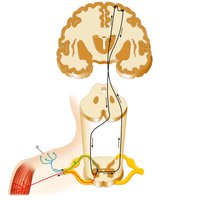
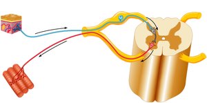
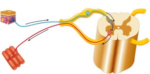
The nervous system processes information through a sequence of sensory input, integration, and motor output. Sensory receptors detect changes, interneurons process the information, and motor neurons initiate a response.
Sensory Input: Sensory receptors monitor internal and external changes and send gathered stimuli to the CNS for processing (afferent pathway).
Integration: The CNS processes information from multiple receptors and dictates an appropriate response.
Motor Output: Initiation and activation of a motor response (efferent pathway).
Example: A fly lands on your arm, activating sensory neurons. The brain processes this input and activates muscles to brush off the fly.
Neurons and Supporting Cells
Types of Cells in the Nervous System
The nervous system contains two main groups of cells: neurons and supporting (glial) cells.
Neurons: Excitable cells that respond to stimuli and conduct electrical impulses. They allow communication between the CNS and the rest of the body, can survive for over 100 years, and have a high metabolic rate requiring abundant oxygen and glucose.
Supporting Cells (Glia): Non-excitable cells that help neurons function more efficiently by providing support, insulation, and protection.
Structure of a Typical Neuron
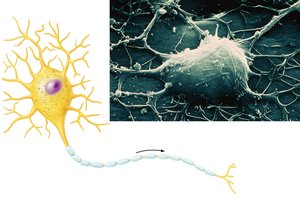
Neurons have specialized structures for receiving, processing, and transmitting information.
Cell Body (Soma): Contains the nucleus and organelles.
Dendrites: Receptive regions that carry information toward the cell body. Neurons may have none to multiple dendrites.
Axon: A single, long process that carries information away from the cell body to axon terminals, where it forms synapses with other cells. Axons may be surrounded by a myelin sheath for faster conduction.
Myelin Sheaths
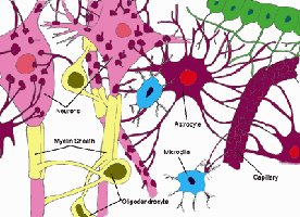
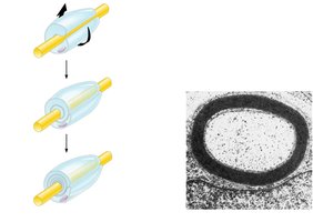
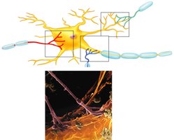
Myelin sheaths are insulating layers that surround axons, increasing the speed of action potential conduction. In the PNS, Schwann cells form the myelin sheath, while in the CNS, oligodendrocytes perform this function.
Myelinated Axons: Schwann cells wrap around a single axon, forming a thick sheath for rapid conduction.
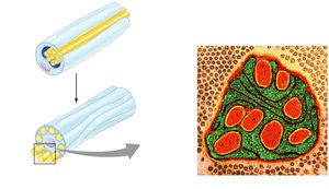
Unmyelinated Axons: Schwann cells surround multiple axons without forming a thick sheath, resulting in slower conduction.
Myelin Sheath in the CNS
In the CNS, oligodendrocytes extend multiple processes to wrap around axons of different interneurons, forming the myelin sheath.
Multiple Sclerosis
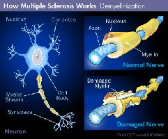
Multiple sclerosis (MS) is a disease characterized by the gradual destruction of myelin in the CNS, leading to the formation of plaques and disruption of action potential conduction. This results in progressive motor and sensory problems.
Classification of Neurons
Structural Classification
Multipolar Neurons: Most common type; have one axon and multiple dendrites. Found as motor neurons and interneurons.
Bipolar Neurons: Have one axon and one dendrite; found in special sensory organs (e.g., retina of the eye).
Unipolar (Pseudo-unipolar) Neurons: Have a single process that splits into two branches; typical of sensory neurons.
Functional Classification
Sensory (Afferent) Neurons: Transmit impulses toward the CNS; cell bodies are clustered in ganglia and are usually unipolar.
Motor (Efferent) Neurons: Transmit impulses away from the CNS to effectors (muscles and glands); cell bodies are within the CNS and are multipolar.
Interneurons (Association Neurons): Located entirely within the CNS; process information and are the most numerous type (99% of all neurons).
Neuron vs. Nerve
Neuron: A single excitable cell that carries electrical impulses.
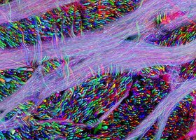
Nerve: A collection of many axons (nerve fibers) in the PNS, containing both motor and sensory fibers.
Tract: A collection of axons in the CNS (interneurons).
Structure of a Nerve
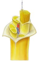
Nerves are organs composed of axons, Schwann cells, connective tissue, and blood vessels. They are organized into bundles called fascicles, each surrounded by connective tissue layers:
Endoneurium: Surrounds individual axons (thin reticular connective tissue).
Perineurium: Surrounds each fascicle of axons (fibrous connective tissue).
Epineurium: Surrounds the entire nerve (dense irregular connective tissue).
Types of Nerves
Mixed Nerves: Contain both sensory and motor neurons (most common type).
Sensory Nerves: Contain only sensory neurons (rare).
Motor Nerves: Contain only motor neurons (rare).
Peripheral Nervous System: Somatic and Visceral Divisions
Somatic vs. Visceral Functions
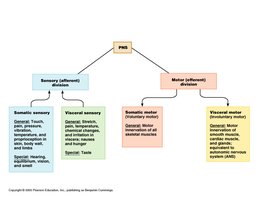
The PNS is divided into somatic and visceral divisions, each with sensory and motor components:
Somatic Functions: Involve structures such as muscles and skin.
Visceral Functions: Involve internal organs (viscera) within body cavities, such as the liver, stomach, heart, blood vessels, and lungs.
Synapses
Structure and Function of Synapses
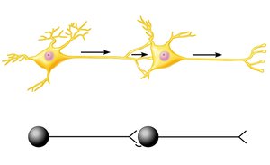
A synapse is the site where neurons communicate with each other or with other types of cells. Communication occurs via the release of neurotransmitters from the presynaptic neuron into the synaptic cleft, which then bind to receptors on the postsynaptic neuron, initiating a response.
Presynaptic Neuron: Carries the impulse toward the synapse.
Postsynaptic Neuron: Carries the impulse away from the synapse.
Neurotransmitters: Chemical messengers (e.g., acetylcholine, norepinephrine) released into the synaptic cleft.
Types of Synapses
Axodendritic: Presynaptic axon to postsynaptic dendrite (most common).
Axosomatic: Presynaptic axon to postsynaptic cell body.
Axoaxonic: Presynaptic axon to postsynaptic axon.
Supporting Cells (Glia) of the Nervous System
Central Nervous System Glia
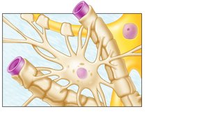
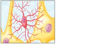
Astrocytes: Most abundant; surround capillaries and neurons, nourish neurons, and regulate neurotransmitter levels.
Microglia: Smallest and least abundant; act as macrophages to remove pathogens and debris.
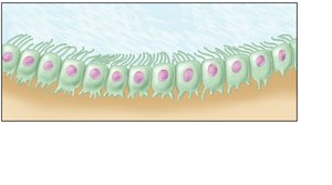
Ependymal Cells: Line the ventricles of the brain and central canal of the spinal cord; cilia aid in circulating cerebrospinal fluid (CSF).
Oligodendrocytes: Form the myelin sheath around CNS axons.
Peripheral Nervous System Glia
Schwann Cells: Form myelin sheath around axons in the PNS, allowing for faster conduction of nerve impulses.
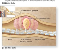
Satellite Cells: Surround neuron cell bodies in ganglia, providing insulation and preventing information crossover.
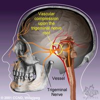
Clinical Correlation: Tic Douloureux (Trigeminal Neuralgia)
Tic Douloureux is a nerve disorder causing severe facial pain. It is often associated with degeneration of satellite cells surrounding the cell bodies of sensory neurons in the trigeminal nerve, leading to abnormal activation of pain pathways.