 Indietro
Joints: Structure, Types, and Cartilage in the Human Skeleton
Guida di studio - Note intelligenti
 Appunti personalizzati basati sui tuoi materiali, ampliati con definizioni chiave, esempi e contesto.
Joint Composition and Connective Tissues
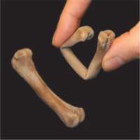
Bone Matrix and Composition
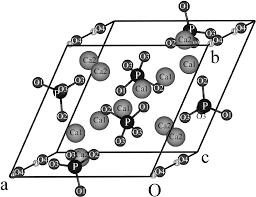
The bone matrix is a composite material consisting of both organic and inorganic components, which together provide strength and resilience to bone tissue.
Inorganic Component: Primarily hydroxyapatite (~60%), which is hard and resists compression.
Organic Component: Mostly type I collagen (~30%), which is strong and resists tension.
Water: Makes up about 10% of the matrix.
Hydroxyapatite alone: Hard but brittle.
Collagen alone: Strong but deformable.
Combination: Provides a strong, flexible, and shatter-resistant structure.
Dense Connective Tissues
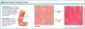
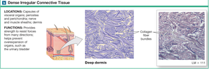
Dense connective tissues are critical for transmitting forces and providing structural support in the body. They are classified as regular or irregular based on fiber arrangement.
Dense Regular Connective Tissue: Collagen fibers are tightly packed and parallel, transmitting force in one direction. Found in tendons and ligaments.
Dense Irregular Connective Tissue: Collagen fibers are interwoven with no consistent pattern, providing support for areas with multidirectional stresses. Found in dermis, organ capsules, and sheaths around cartilage and bone.
Terminology of Connective Tissues
Fibrous: Refers to dense regular or irregular connective tissue.
Tendon: Dense regular CT connecting muscle to bone (M2B), cord-like.
Aponeurosis: Dense regular CT connecting muscle to bone or muscle to muscle (M2B or M2M), sheet-like.
Ligament: Dense regular CT connecting bone to bone (B2B), cord-like or band-like.
Interosseous membrane: Dense regular CT connecting bone to bone (B2B), sheet-like.
Cartilage: Structure, Properties, and Types
Cartilage Development and Properties
Cartilage is a firm but flexible supporting connective tissue produced by chondrocytes. It forms the template for bone development and is essential in joint function.
Cartilage Matrix: Gel-like, composed of collagen and proteoglycans.
Viscoelasticity: Cartilage can deform under force and gradually return to its original shape.
Avascular and Aneural: Cartilage lacks blood vessels and nerves, limiting its repair capacity and thickness.
Perichondrium: Dense irregular CT sheath containing nerves and blood vessels, surrounds most cartilage except articular cartilage.
Comparison of Cartilage and Bone
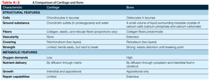
Cartilage and bone differ in structure, function, and repair capabilities. The following table summarizes these differences:
Characteristic | Cartilage | Bone |
|---|---|---|
Cells | Chondrocytes in lacunae | Osteocytes in lacunae |
Ground Substance | Chondroitin sulfate in proteoglycans and water | Small volume of liquid surrounding insoluble crystals of calcium salts (calcium phosphate and calcium carbonate) |
Fibers | Collagen, elastic, and reticular fibers (proportions vary) | Collagen fibers predominate |
Covering | Perichondrium (except articular cartilage) | Periosteum |
Vascularity | Avascular | Vascular |
Oxygen Demands | Low | High |
Nutrient Delivery | By diffusion through matrix | By diffusion through cytoplasm and interstitial fluid in canaliculi |
Growth | Interstitial and appositional | Appositional only |
Repair Capabilities | Limited | Extensive |
Cartilage Subtypes
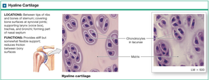
Hyaline Cartilage
Hyaline cartilage is the most common type, providing flexible support and reducing friction between bony surfaces.
Structure: Collagen is tightly packed but not bundled; high ground substance content makes it springy and shock-absorbing.
Perichondrium: Present except at articular surfaces.
Location: Ends of ribs, bones of sternum, covering bone surfaces at synovial joints, supporting larynx, trachea, bronchi, and forming part of nasal septum.
Elastic Cartilage
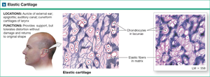
Elastic cartilage is similar to hyaline but contains more elastic fibers, making it very flexible.
Structure: Contains a perichondrium and abundant elastic fibers.
Location: Auricle of external ear, epiglottis, auditory canal, cuneiform cartilages of larynx.
Function: Provides support but tolerates distortion without damage and returns to original shape.
Fibrocartilage
Fibrocartilage is tough and load-bearing, with tightly packed and interwoven collagen fibers and low ground substance.
No Perichondrium: Lacks blood vessels, relies on diffusion for nutrients and oxygen.
Location: Intervertebral discs, pubic symphysis, menisci of knee.
Function: Absorbs shock and enables slight movement between bones.
Axial and Appendicular Joints
Axial Skeleton Joints
The axial skeleton includes joints such as those between ribs and sternum, vertebrae, and skull sutures.
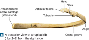
Costal Cartilage: Connects ribs to sternum, forming chondrocostal and sternocostal joints.
Intervertebral Discs: Fibrocartilaginous cushions between vertebrae, absorb shocks and allow movement.
Appendicular Skeleton Joints
The appendicular skeleton consists of bilateral bones in the limbs, with shared homology between upper and lower limbs.
Carpus (Wrist): Contains 8 carpals, 5 metacarpals, and 14 phalanges.
Wrist Joint: Articulations between radius and proximal carpal bones; ulna contacts via a fibrocartilaginous articular disc.
Pelvic Girdle: Connects via fibrocartilage (pubic symphysis).
Pectoral Girdle: Connects bone-to-bone via sternum.
Joint Types and Functional Designations
Functional Classification of Joints
Joints are classified by their degree of movement:
Synarthrosis: Immovable; connection can be fibrous, cartilaginous, or ossified.
Amphiarthrosis: Slightly movable; connection can be fibrous or cartilaginous.
Diarthrosis (Synovial): Freely movable; indirect connection via articular cartilage, fibrous capsule, and synovial fluid-filled cavity.
Structural Classification of Joints
Fibrous Joints: Bones joined by dense connective tissue (e.g., sutures, syndesmosis, gomphosis).
Cartilaginous Joints: Bones joined by cartilage (e.g., synchondrosis, symphysis).
Synovial Joints: Bones separated by a fluid-filled cavity, allowing free movement.
Synovial Joints: Structure and Accessory Features
Synovial Membranes and Fluid
Highly mobile joints are enclosed by a fibrous capsule containing a synovial membrane, which produces synovial fluid.
Synovial Fluid: Lubricant with nutrients, absorbs and disperses shock, viscoelastic, 'yolk-like' consistency.
Synoviocytes: Embedded in areolar connective tissue, produce synovial fluid.
Accessory Structures of Synovial Joints
Fat Pads: Protect and pack the joint.
Meniscus: Fibrocartilage pad that improves joint fit, shock absorption, and stability.
Bursa: Fluid cushions that reduce friction and absorb shock where tendons rub on bones.
Ligaments: Dense regular CT connecting bone to bone; avascular and difficult to heal.
Tendons: Connect muscle to bone and help stabilize joints.
Synovial Joint Movements
Types of Synovial Joint Movements
Plane Joints: Allow gliding movements.
Hinge Joints: Permit flexion and extension.
Pivot Joints: Allow rotation (e.g., pronation/supination).
Condylar Joints: Permit movement in two planes.
Saddle Joints: Each side has convex and concave surfaces; allows greater range of motion (e.g., thumb).
Ball-and-Socket Joints: Allow movement in multiple axes and planes.
Summary Table: Joint Types and Features
Joint Type | Structure | Movement | Example |
|---|---|---|---|
Synarthrosis | Fibrous, cartilaginous, or ossified | Immovable | Sutures, gomphosis |
Amphiarthrosis | Fibrous or cartilaginous | Slightly movable | Pubic symphysis, intervertebral discs |
Diarthrosis (Synovial) | Articular cartilage, capsule, synovial fluid | Freely movable | Shoulder, hip, knee |
Key Equations and Concepts
Bone Matrix Equation:
Viscoelasticity: (for linear elastic materials)
Conclusion
Understanding the composition, structure, and function of joints and their associated connective tissues is fundamental to the study of anatomy and physiology. The interplay between bone, cartilage, and connective tissue determines the range of motion, stability, and repair capacity of the skeletal system.
