 Indietro
Lower Digestive Path: Structure, Function, and Regulation (Module 7: Part 3)
Guida di studio - Note intelligenti
 Appunti personalizzati basati sui tuoi materiali, ampliati con definizioni chiave, esempi e contesto.
Lower Digestive Path
Small Intestine
The small intestine is the primary site for nutrient absorption in the digestive system. It consists of three main regions: the duodenum, jejunum, and ileum. Each region plays a distinct role in digestion and absorption.
Duodenum: Receives chyme from the stomach and digestive enzymes from the pancreas and liver.
Jejunum: Major site for chemical digestion and nutrient absorption.
Ileum: Absorbs remaining nutrients and connects to the large intestine.
Lymphatic tissue: Protects against pathogens entering with food.
Intestinal Structure and Cells
The small intestine contains specialized structures and cells to maximize absorption and protect the body.
Intestinal villi: Finger-like projections that increase surface area for absorption.
Microvilli: Tiny extensions on absorptive cells, further increasing surface area.
Absorptive cells: Take up nutrients from the lumen.
Goblet cells: Secrete mucus to protect and lubricate the intestinal lining.
Intestinal glands: Produce intestinal juice containing digestive enzymes.
Paneth cells: Secrete antimicrobial substances.
Enteroendocrine cells: Release hormones regulating digestion.
Vessels & lacteal: Transport absorbed nutrients (lacteals carry fats).
Digestion in the Small Intestine
Digestion in the small intestine involves both brush border enzymes (attached to microvilli) and enzymes in the lumen.
Carbohydrates: Broken down by amylase and brush border enzymes into monosaccharides.
Proteins: Digested by proteases and peptidases into amino acids.
Lipids: Emulsified by bile salts and digested by lipases into fatty acids and monoglycerides.
Nucleic acids: Broken down by nucleases into nucleotides.
Absorption in the Small Intestine
Absorption occurs through various mechanisms, including active transport, diffusion, and facilitated diffusion. Most nutrients, water, and electrolytes are absorbed here.
Pancreas
Structure and Function
The pancreas is both an endocrine and exocrine organ, crucial for digestion and metabolic regulation.
Pancreatic islets: Secrete insulin and glucagon to regulate blood glucose.
Pancreatic acini: Produce pancreatic juice containing digestive enzymes.
Pancreatic juice: Contains bicarbonate (neutralizes stomach acid), amylase (digests carbohydrates), lipase (digests fats), nucleases (digest nucleic acids), proteases and peptidases (digest proteins), and proenzymes (inactive forms activated in the intestine).
Liver
Structure
The liver is organized into lobules, each containing a central vein and surrounded by interlobular septa. Kupffer cells (macrophages) line the sinusoids, and bile canaliculi collect bile produced by hepatocytes.
Lobules: Functional units of the liver.
Central vein: Drains blood from each lobule.
Kupffer cells: Remove debris and pathogens.
Bile canaliculi: Collect bile for transport to hepatic ducts.
Hepatic ducts, cystic duct, common bile duct: Pathways for bile flow.
Functions
The liver performs metabolic and hematological regulation, stores nutrients, removes waste, inactivates drugs, and produces bile for fat emulsification.
Metabolic regulation: Extracts and stores nutrients, removes waste, stores vitamins and minerals.
Hematological regulation: Filters blood, removes old cells, produces plasma proteins.
Bile production: Bile salts emulsify fats, aiding digestion.
Blood Flow Through the Liver
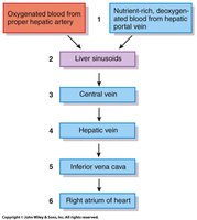
Blood enters the liver from the hepatic artery (oxygenated) and hepatic portal vein (nutrient-rich, deoxygenated). It flows through sinusoids, central vein, hepatic vein, and finally to the heart.
Large Intestine
Structure
The large intestine includes the cecum, ileocecal valve, appendix, colon, rectum, and anal canal. Sphincters control passage and elimination of waste.
Cecum: Receives material from the ileum.
Ileocecal valve: Regulates flow from small to large intestine.
Appendix: Contains lymphatic tissue.
Colon: Main site for water and vitamin absorption.
Rectum and anal canal: Store and eliminate feces.
Sphincters: Control defecation.
Functions
Reabsorption of water: Prevents dehydration.
Absorption of vitamins: Especially vitamin K, biotin, and B5 produced by gut bacteria.
Storage of feces: Prepares waste for elimination.
Control of Digestion
Nervous System Regulation
Digestion is regulated by the central nervous system (CNS), enteric nervous system (ENS), and autonomic nervous system (ANS). The ENS is a complex network within the gut wall, capable of independent function.
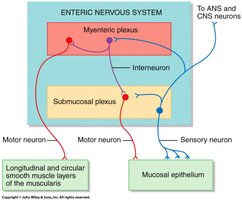
CNS: Coordinates overall digestive activity.
ENS: Controls local gut motility and secretion.
ANS: Modulates digestive activity via sympathetic and parasympathetic input.
Endocrine System Regulation
Hormones released by enteroendocrine cells and other organs regulate digestive processes.
CCK (Cholecystokinin): Stimulates bile and pancreatic enzyme release.
ACTH (Adrenocorticotropic hormone): Indirectly affects metabolism.
Leptin: Regulates appetite and energy balance.
Ghrelin: Stimulates hunger.
Digestive Regulation Phases
Cephalic Phase
The cephalic phase is triggered by the sight, smell, or thought of food, stimulating gastric secretions via neural pathways.
Gastric Phase
The gastric phase begins when food enters the stomach, activating neural and hormonal responses to increase gastric motility and secretion.
Intestinal Phase
The intestinal phase starts as chyme enters the small intestine, with neural and hormonal mechanisms regulating further digestion and absorption.
Control of Hunger
Hunger regulation is complex and involves nervous system stretch receptors, hormones, and psychological/social factors.
Stretch receptors: Signal fullness.
Hormones: CCK, leptin, ghrelin influence appetite.
Psychological/social factors: Affect eating behavior.
