 Indietro
Neurophysiology: Structure and Function of Neurons and Neuroglia
Guida di studio - Note intelligenti
 Appunti personalizzati basati sui tuoi materiali, ampliati con definizioni chiave, esempi e contesto.
Neuroglia and Neuron Structure
Types of Neuroglia in the Central and Peripheral Nervous Systems
Neurons are highly specialized cells that require support from neuroglia, which are non-neuronal cells that maintain homeostasis, form myelin, and provide support and protection for neurons. There are six main types of neuroglia: four in the central nervous system (CNS) and two in the peripheral nervous system (PNS).
Astrocytes (CNS): Star-shaped, most abundant; anchor neurons to capillaries, facilitate nutrient exchange, guide migration of young neurons, and clean up excess neurotransmitters and ions.
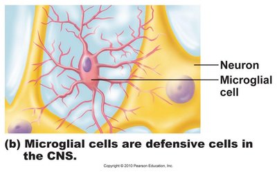
Microglia (CNS): Defensive cells; monitor neuron health and can transform into macrophages to engulf microorganisms and debris.
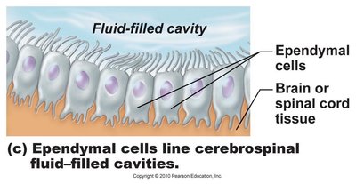
Ependymal cells (CNS): Line cavities of the brain and spinal cord; create a barrier between cerebrospinal fluid (CSF) and CNS tissue, and their cilia circulate CSF.
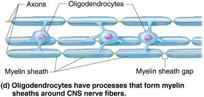
Oligodendrocytes (CNS): Form myelin sheaths around CNS neurons, increasing the speed of electrical transmission.
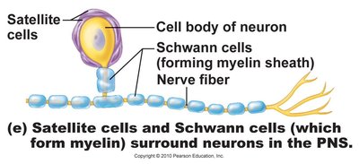
Satellite cells (PNS): Surround neuron cell bodies in ganglia; functionally similar to astrocytes.
Schwann cells (PNS): Form myelin sheaths around larger PNS neurons; essential for peripheral nerve regeneration.

Classification of Neurons
Structural and Functional Classification
Neurons are classified based on the number of processes extending from the cell body (structural) and the direction of nerve impulse transmission (functional).
Structural: Unipolar, bipolar, and multipolar neurons.
Functional:
Sensory (afferent): Transmit impulses toward the CNS; primary sensory neurons are mostly unipolar.
Motor (efferent): Transmit impulses away from the CNS to effectors; multipolar, most cell bodies reside in CNS.
Association (interneurons): Integrate information between sensory and motor neurons; multipolar, mostly within CNS.
Electrical Activity of Neurons
Resting Membrane Potential (RMP)
The resting membrane potential is the voltage difference across the cell membrane when the neuron is not transmitting a signal. It is negative inside relative to outside, primarily due to differential permeability to Na+ and K+ ions and the activity of the Na+/K+ pump.
Key Points:
K+ is primarily inside cells; Na+ is primarily outside.
Membrane is more permeable to K+ than Na+ at rest.
Na+/K+ pump moves 3 Na+ out for every 2 K+ in.
Equation:
Types of Ion Channels
Channels in the plasma membrane allow ions to move passively in response to electrochemical gradients. There are two main types:
Leakage channels: Always open.
Gated channels: Require a signal to open/close; can be chemically-gated or voltage-gated.
Depolarization vs. Hyperpolarization
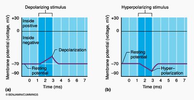
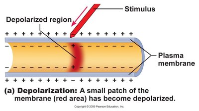
Depolarization increases the probability of producing nerve impulses, while hyperpolarization decreases it.
Graded Potentials
Characteristics and Types
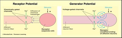
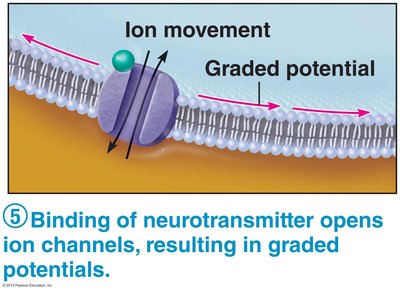
Graded potentials are short-lived changes in membrane potential, produced in dendrites, and their magnitude depends on the strength of the stimulus. They decrease with distance traveled and can be depolarizations or hyperpolarizations.
Generator/Receptor Potentials: Triggered by external stimuli or neurotransmitter release.
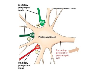
Postsynaptic Potentials: Triggered by neurotransmitter binding.
Action Potentials
Definition and Phases
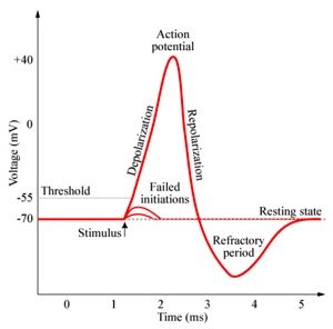
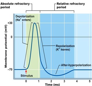
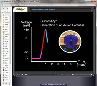
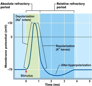
An action potential (AP) is a brief reversal of membrane potential, typically from -70 mV to +30 mV. Only axons generate APs, which do not decrease in amplitude with distance.
Phases:
Depolarization: Voltage-gated Na+ channels open.
Repolarization: Na+ channels close, K+ channels open.
After-hyperpolarization: K+ channels remain open briefly.
Threshold: The minimum membrane potential required to trigger an AP (typically -55 to -50 mV).
All-or-None Principle: APs are always the same size; stimulus intensity is coded by frequency.
Equation:
Propagation of Action Potentials
Mechanism and Myelination
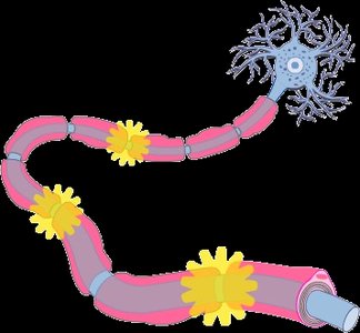
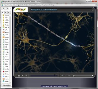
Action potentials propagate along the axon, not by conduction but by sequential opening of voltage-gated channels. Propagation is unidirectional due to refractory periods. Myelin sheaths, produced by Schwann cells (PNS) and oligodendrocytes (CNS), act as insulators and increase the speed of impulse propagation via saltatory conduction at nodes of Ranvier.
Synaptic Transmission
Types of Synapses and Mechanisms
Synapses are junctions between two neurons or a neuron and an effector. There are two types: electrical (direct current flow via gap junctions) and chemical (neurotransmitter-mediated).
Electrical Synapses: Rapid, synchronized transmission; found in embryonic tissue, eye movement, cardiac, and smooth muscle.
Chemical Synapses: Neurotransmitters open or close ion channels, affecting membrane permeability and potential.
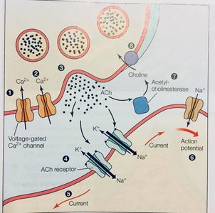
Mechanism of Chemical Synaptic Transmission:
Ca2+ gates open in presynaptic terminal.
Neurotransmitter release into synaptic cleft.
Neurotransmitter binds to postsynaptic receptors.
Ion channels open in postsynaptic membrane, causing depolarization or hyperpolarization.
Termination and Synaptic Delay
Termination: Neurotransmitter can be degraded by enzymes, reuptaken by presynaptic terminal, or diffuse away.
Synaptic Delay: Slowest step in neurotransmission; takes 0.3–5 ms for neurotransmitter release, diffusion, and binding.
Postsynaptic Potentials and Summation
EPSPs and IPSPs
Postsynaptic potentials are graded and occur in the dendritic region. Excitatory postsynaptic potentials (EPSPs) cause depolarization, while inhibitory postsynaptic potentials (IPSPs) cause hyperpolarization.
Summation: Multiple EPSPs and IPSPs can be summed temporally or spatially at the axon hillock to determine whether an AP is generated.
Neurotransmitter Classification
Chemical and Functional Classification
Chemical: Acetylcholine, biogenic amines (dopamine, norepinephrine, epinephrine, serotonin, histamine), amino acids (glutamate, aspartate, glycine, GABA), neuropeptides (substance P, endorphins), purines (ATP, adenosine), gases & lipids (NO, CO, H2S), endocannabinoids.
Functional: Excitatory vs. inhibitory effects; direct vs. indirect actions (second messenger systems).
Neural Circuits
Organization and Processing
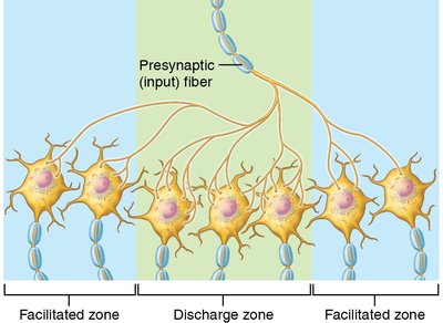
Neural processing can be serial (reflexes) or parallel (complex behaviors). Neurons in the CNS are organized into pools to handle information processing.
Additional info: These notes cover Chapter 11: Fundamentals of the Nervous System and Nervous Tissue, focusing on neuroglia, neuron structure, membrane potentials, action potentials, synaptic transmission, and neural circuits.
