 Indietro
Osseous Tissue and Bone Structure: Study Notes for Anatomy & Physiology
Guida di studio - Note intelligenti
 Appunti personalizzati basati sui tuoi materiali, ampliati con definizioni chiave, esempi e contesto.
Osseous Tissue and Bone Structure
Divisions and Functions of the Skeletal System
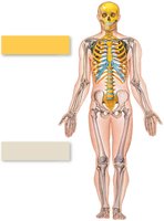
The skeletal system is divided into two major regions: the axial skeleton and the appendicular skeleton. Each division plays a distinct role in supporting and facilitating movement in the human body.
Axial Skeleton: Composed of bones along the longitudinal axis (skull, thorax, vertebral column).
Appendicular Skeleton: Includes bones of the limbs and girdles (pectoral and pelvic) that attach limbs to the axial skeleton.
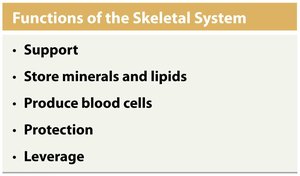
The skeletal system serves several essential functions:
Support: Provides a framework for the body.
Storage of Minerals: Stores calcium and phosphate, releasing them to maintain homeostasis.
Blood Cell Production: Red bone marrow produces red and white blood cells, and platelets.
Protection: Shields vital organs (e.g., skull, ribs, vertebral column, pelvis).
Leverage: Bones act as levers for muscle action, enabling movement.
Classification of Bones by Shape and Structure
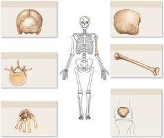
Bones are classified based on their shape and internal structure, which reflects their function and location in the body.
Flat Bones: Two parallel surfaces; protect underlying structures (e.g., skull, ribs).
Sutural (Wormian) Bones: Small bones within cranial sutures.
Long Bones: Long and slender (e.g., humerus, femur).
Irregular Bones: Complex shapes with projections (e.g., vertebrae).
Sesamoid Bones: Small, embedded in tendons (e.g., patella).
Short Bones: Cube-shaped (e.g., carpal bones).
Surface Features of Bones
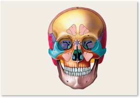
Bones display various surface features that serve as sites for muscle attachment, passage of nerves and blood vessels, and articulation with other bones.
Canal or Meatus: Large passageway through bone.
Process: Any projection or bump.
Sinus: Air-filled chamber within bone.
Foramen: Small rounded hole for vessels and nerves.
Fissure: Elongated cleft or gap.
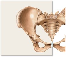
Pelvic bones also have distinct features:
Crest: Prominent ridge.
Fossa: Shallow depression.
Line: Low, delicate ridge.
Spine: Pointed or narrow process.
Ramus: Extension making an angle with the rest of the bone.
Structure of a Typical Long Bone
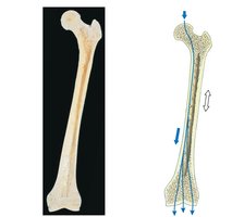
Long bones have specialized regions and internal structures that support their function and growth.
Epiphysis: Ends of the bone.
Metaphysis: Region where shaft joins epiphyses; site of growth at the epiphyseal plate.
Diaphysis: Shaft or elongated body.
Articular Cartilage: Covers joint surfaces; limited repair capacity.
Medullary (Marrow) Cavity: Contains yellow marrow (fat).
Periosteum: Membrane covering bone surface.
Types of bone tissue:
Compact Bone: External layer; strong and dense.
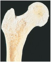
Spongy Bone: Internal; contains spaces for red marrow, lighter and oriented along stress lines.
Blood Supply to Bone
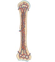
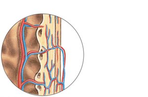
Bones have an extensive blood supply, essential for growth, repair, and metabolic activity.
Nutrient Artery and Vein: Enter through nutrient foramen, supply diaphysis.
Epiphyseal and Metaphyseal Arteries/Veins: Supply epiphysis and metaphysis.
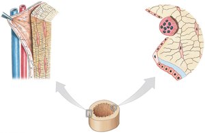
Periosteal Arteries/Veins: Supply periosteum and outer compact bone.
Sensory Nerves: Accompany blood vessels, providing sensation.
Cell Types in Bone and Bone Matrix
Bone tissue contains several specialized cell types, each with distinct functions in bone formation, maintenance, and remodeling.
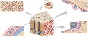
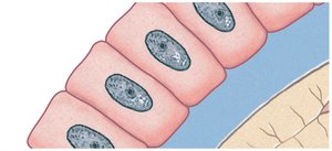
Osteoprogenitor Cells: Stem cells that differentiate into osteoblasts.
Osteoblasts: Bone-forming cells; produce osteoid and initiate calcification.
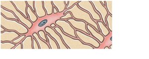
Osteocytes: Mature bone cells; maintain bone matrix, reside in lacunae, connected by canaliculi.
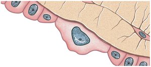
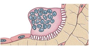
Osteoclasts: Bone-resorbing cells; break down bone matrix (osteolysis).
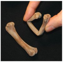
The bone matrix is primarily composed of calcium salts (providing hardness) and collagen fibers (providing tensile strength).
Structure and Function of Compact and Spongy Bone
Compact and spongy bone differ in their structure and function, reflecting their roles in the skeleton.
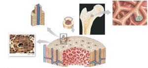
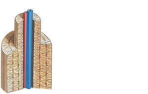
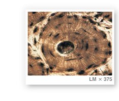
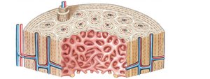
Compact Bone: Composed of osteons (cylindrical units), central canals, concentric lamellae, lacunae, canaliculi, perforating canals, interstitial lamellae, and circumferential lamellae.
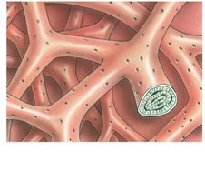
Spongy Bone: Contains trabeculae (struts and plates), oriented along stress lines, lighter, and allows diffusion of nutrients through canaliculi.
Appositional Bone Growth
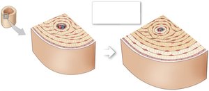
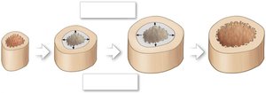
Bone increases in diameter through appositional growth, involving the periosteum and endosteum.
Periosteum: Contains osteoblasts that add layers to the outer surface.
Endosteum: Contains osteoclasts and osteoblasts for remodeling.
Process: Osteoblasts add circumferential lamellae; osteoclasts enlarge the medullary cavity.
Endochondral Ossification and Bone Growth in Length
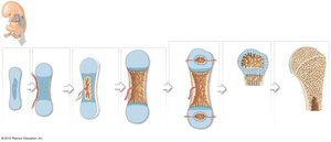
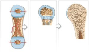
Most bones develop through endochondral ossification, where bone forms inside hyaline cartilage.
Process: Chondrocytes die, osteoblasts form bone collar, blood vessels invade, primary and secondary ossification centers form, cartilage remains at articular surfaces and epiphyseal plate.
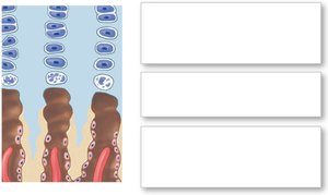
Growth in Length: Occurs at epiphyseal plate; cartilage cells divide and are replaced by bone. At puberty, increased hormones accelerate bone formation, leading to epiphyseal closure.
Intramembranous Ossification
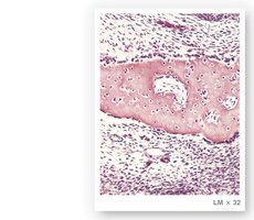
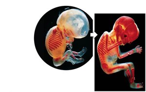
Intramembranous ossification forms bone directly from mesenchymal tissue, without a cartilage model. It is responsible for the development of flat bones of the skull, clavicle, and mandible.
Process: Mesenchymal cells differentiate into osteoblasts at ossification centers, secrete matrix, become osteocytes, matrix forms spongy bone, periosteum forms, and superficial layers become compact bone.
Timing: Begins around the 8th week of development; by 16 weeks, most adult skeleton is identifiable.
Abnormalities of Bone Formation and Growth
Various endocrine and metabolic disorders can affect bone growth and development.
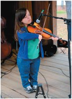
Pituitary Growth Failure: Reduced growth hormone leads to short bones.
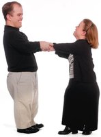
Achondroplasia: Early closure of epiphyseal plates results in short, stocky limbs.
Gigantism: Excess growth hormone before puberty causes excessive bone lengthening.
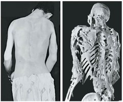
Acromegaly: Excess growth hormone after epiphyseal closure causes bone thickening.


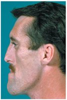
Regulation of Blood Calcium Levels
Calcium is the most abundant mineral in the body, and its levels are tightly regulated by several mechanisms involving the intestines, kidneys, and bone cells.
Absorption: Dietary calcium absorbed in intestines (hormonally regulated).
Loss: Calcium lost in urine (hormonally regulated).
Bone Remodeling: Osteoblasts deposit calcium; osteoclasts resorb bone, releasing calcium.
Hormonal Regulation of Calcium Homeostasis
Blood calcium levels are maintained by the actions of parathyroid hormone (PTH) and calcitonin.
Low Ca2+: PTH increases osteoclast activity, stimulates calcitriol production, increases intestinal absorption, and reduces urinary loss.
High Ca2+: Calcitonin inhibits osteoclasts, increases calcium deposition in bone, and promotes urinary excretion.
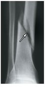
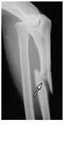
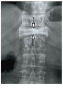
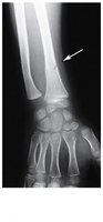
Types of Fractures and Fracture Healing
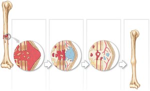
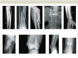
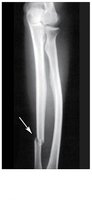
Fractures are classified by their pattern and severity. Healing involves several stages, provided blood supply and bone membranes survive.
Closed (Simple) Fracture: Bone breaks but does not protrude through skin.
Open (Compound) Fracture: Bone protrudes through skin, risk of infection.
Transverse: Break across long axis.
Spiral: Twisting stress.
Displaced: Abnormal alignment.
Compression: Vertebrae crushed.
Greenstick: One side breaks, other bends (children).
Comminuted: Bone fragments into pieces.
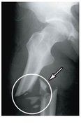
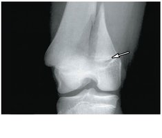
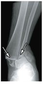
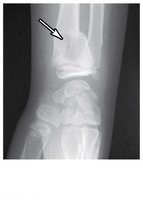
Epiphyseal: Along growth plate.
Pott’s: Ankle, both leg bones.
Colles: Distal radius, fall injury.
Medical Terminology
Osteomyelitis: Infection of bone, often caused by Staphylococcal bacteria, characterized by fever, pain, and inflammation.
Osteoporosis: Decreased bone mass and strength, often due to hormonal changes; bone resorption exceeds formation, increasing fracture risk.
Additional info: The notes above expand on brief points with academic context, definitions, and examples, and include only images that are directly relevant to the explanation of each paragraph.
