 Indietro
Peripheral Nervous System: Structure, Function, and Clinical Relevance
Guida di studio - Note intelligenti
 Appunti personalizzati basati sui tuoi materiali, ampliati con definizioni chiave, esempi e contesto.
Peripheral Nervous System (PNS)
Overview and Functional Organization
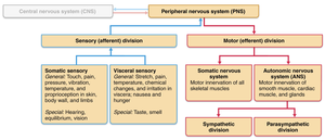
The Peripheral Nervous System (PNS) consists of all nervous structures outside the brain and spinal cord. It connects the central nervous system (CNS) to the rest of the body, facilitating communication between sensory receptors, muscles, glands, and the CNS. The PNS is functionally divided into sensory (afferent) and motor (efferent) divisions.
Sensory Division: Carries information from sensory receptors to the CNS.
Motor Division: Transmits commands from the CNS to effector organs (muscles and glands).
Structures: Sensory receptors, nerves (bundles of axons), ganglia (clusters of neuronal cell bodies), and motor endings.
Sensory Receptors
Classification and Function
Sensory receptors are specialized structures that detect stimuli and initiate nerve impulses. They are classified by location and stimulus type:
By Location:
Exteroreceptors: Detect stimuli outside the body (e.g., touch, temperature).
Interoreceptors: Detect internal stimuli (e.g., visceral pain, chemical changes).
Proprioreceptors: Located in muscles and bones; monitor body position and movement.
By Stimulus Type:
Mechanoreceptors: Respond to touch, pressure, vibration.
Thermoreceptors: Detect temperature changes.
Chemoreceptors: Sense chemical stimuli.
Photoreceptors: Respond to light.
Nociceptors: Detect pain.
General Sensory Receptors
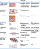
General sensory receptors are distributed throughout the body and include free nerve endings and encapsulated nerve endings. They play roles in detecting touch, pressure, vibration, and proprioception.
Free Nerve Endings: Abundant in epithelia and dermis; detect itch and light touch.
Encapsulated Nerve Endings: Mechanoreceptors with connective tissue capsules. Types include:
Tactile corpuscles (Meisner’s): Fine touch, found in hairless skin.
Lamellar corpuscles (Pacinian): Deep pressure and vibration.
Bulbous corpuscles (Ruffini): Continuous pressure.
Proprioreceptors:
Muscle spindles: Detect muscle length changes.
Tendon organs: Monitor tendon tension.
Joint kinesthetic receptors: Monitor stretch in synovial joints.
Cranial Nerves
Structure and Function
There are 12 pairs of cranial nerves, each with specific sensory, motor, or mixed functions. The first two attach to the forebrain; the rest to the brainstem.
I – Olfactory: Smell (sensory)
II – Optic: Vision (sensory)
III – Oculomotor: Eye movement (motor)
IV – Trochlear: Eye movement (motor)
V – Trigeminal: Facial sensation, chewing (mixed)
VI – Abducens: Lateral eye movement (motor)
VII – Facial: Facial expression (mixed)
VIII – Vestibulocochlear: Hearing and equilibrium (sensory)
IX – Glossopharyngeal: Tongue and pharynx (mixed)
X – Vagus: Visceral organs, diaphragm (mixed)
XI – Accessory: Trapezius and sternocleidomastoid (motor)
XII – Hypoglossal: Tongue (motor)
Mnemonic devices are commonly used to memorize cranial nerve names and functions.
Spinal Nerves
Structure and Distribution
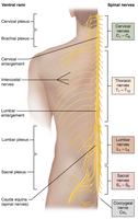
There are 31 pairs of spinal nerves connecting the CNS to muscles, glands, and receptors. Each spinal nerve is associated with a vertebra and is formed from the union of motor and sensory axons.
8 cervical nerves (C1–C8)
12 thoracic nerves (T1–T12)
5 lumbar nerves (L1–L5)
5 sacral nerves (S1–S5)
1 coccygeal nerve (Co)
Spinal Nerve Anatomy
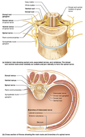
Each spinal nerve is a mixed nerve, connected to the spinal cord by a dorsal root (sensory) and a ventral root (motor). The dorsal root contains sensory neuron cell bodies; the ventral root contains motor neuron axons. Spinal nerves and dorsal root ganglia lie in the intervertebral foramen.
Branches of Spinal Nerves
After exiting the vertebral column, each spinal nerve branches into a dorsal ramus and ventral ramus, both carrying motor and sensory fibers.
Dorsal rami: Innervate deep muscles and skin of the back and neck.
Ventral rami: Innervate muscles and skin of anterior/lateral trunk and limbs.
Roots are either sensory or motor; rami are mixed.
Nerve Plexuses
Structure and Major Plexuses
Nerve plexuses are networks formed by ventral rami (except T1–T12) merging, diverging, and converging. They allow for complex innervation patterns and redundancy.
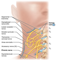
Cervical plexus: Neck; includes phrenic nerve (diaphragm).
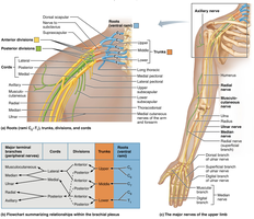
Brachial plexus: Neck and axilla; includes radial, median, ulnar nerves.
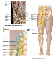
Lumbar plexus: Posterior abdominal wall; includes femoral nerve.
Sacral plexus: Caudal to lumbar plexus; includes sciatic, saphenous, tibial nerves.
Cervical Plexus
Innervates skin and muscles of head and neck, and the diaphragm via the phrenic nerve.
Brachial Plexus
Divided into trunks, divisions, cords, and named nerves. Major nerves include median, ulnar, and radial nerves, which innervate the upper limb.
Lumbar Plexus
The femoral nerve is the largest branch, innervating muscles of the anterior thigh compartment.
Sacral Plexus
The sciatic nerve is the largest branch, supplying most of the lower limb except the anterior and medial thigh. It branches into the tibial and common fibular nerves above the knee.
Dermatomes
Innervation of the Skin
A dermatome is a segment of skin innervated by a single spinal nerve. Dermatomes are clinically important for diagnosing nerve damage and referred pain (e.g., appendicitis pain in the umbilical area, T10).
Disorders of the Peripheral Nervous System
Common Disorders
Shingles (herpes zoster): Viral infection of sensory neurons, often reactivating from childhood chickenpox.
Migraines: Related to sensory innervation of cerebral arteries by the trigeminal nerve; dilation compresses nerve endings causing pain.
Peripheral Neuropathy: Disruption of peripheral nerve function; symptoms include pain, burning, loss of feeling, tingling.
Polio: Viral disease causing paralysis; post-polio syndrome includes muscle/joint pain, weakness, lethargy.
Summary Table: General Sensory Receptors
The following table summarizes the structural class, function, and location of general sensory receptors:
Structural Class | Function/Type | Body Location |
|---|---|---|
Free nerve endings | Mechanoreceptors (itch, touch), thermoreceptors, nociceptors | Most body tissues, especially epithelia and connective tissues |
Modified free nerve endings (tactile discs) | Mechanoreceptors (light touch, slowly adapting) | Basal layer of epidermis |
Hair follicle receptors | Mechanoreceptors (hair deflection, rapidly adapting) | In and surrounding hair follicles |
Tactile corpuscles (Meissner's) | Mechanoreceptors (discriminative touch, rapidly adapting) | Dermal papillae of hairless skin |
Lamellar corpuscles (Pacinian) | Mechanoreceptors (deep pressure, vibration, rapidly adapting) | Dermis, hypodermis, periosteum, mesentery |
Bulbous corpuscles (Ruffini) | Mechanoreceptors (continuous pressure, slowly adapting) | Dermis, hypodermis, joint capsules |
Muscle spindles | Proprioreceptors (muscle stretch) | Skeletal muscles |
Tendon organs | Proprioreceptors (tendon stretch) | Tendons |
Joint kinesthetic receptors | Proprioreceptors (joint stretch) | Joint capsules of synovial joints |
Example: Lamellar corpuscles (Pacinian) are found deep in the skin and respond to vibration and deep pressure, rapidly adapting to changes in stimulus.
Additional info: Dermatomes are mapped clinically to assess sensory loss and referred pain patterns. Nerve plexuses provide redundancy, so damage to one spinal nerve may not result in complete loss of function in a region.
