 Indietro
Renal System Physiology: Nephron Function, Filtration, and Hormonal Regulation (Lab 8 PhysioEx Exercise 9 Review)
Guida di studio - Note intelligenti
 Appunti personalizzati basati sui tuoi materiali, ampliati con definizioni chiave, esempi e contesto.
Renal System Physiology
Nephron Structure and Function
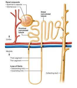
The nephron is the functional unit of the kidney, responsible for filtering blood and forming urine. Each nephron consists of several distinct segments, each with specialized roles in filtration, reabsorption, and secretion.
Renal Corpuscle: Includes the Bowman's capsule and the glomerulus, where blood filtration begins.
Proximal Convoluted Tubule (PCT): Site of major reabsorption of water, ions, and nutrients.
Loop of Henle: Divided into descending and ascending limbs, crucial for creating a concentration gradient in the medulla.
Distal Convoluted Tubule (DCT): Further adjusts the composition of urine.
Collecting Duct: Final site for water reabsorption and urine concentration.
Renal Blood Flow and Filtration
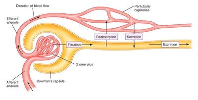
Blood enters the nephron via the afferent arteriole and leaves through the efferent arteriole. Filtration occurs in the glomerulus, where plasma is forced into the Bowman's capsule, initiating urine formation.
Filtration: Movement of water and solutes from blood into the nephron.
Reabsorption: Return of essential substances from the nephron back into the blood.
Secretion: Addition of waste products from blood into the nephron.
Excretion: Removal of urine from the body.
Glomerular Filtration Rate (GFR)
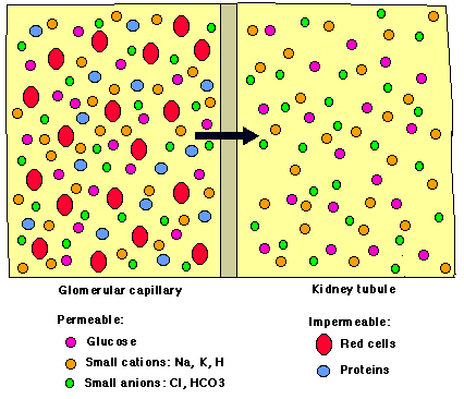
GFR is the rate at which blood is filtered in the glomerulus. It is a key indicator of kidney function, with a normal range of 80–140 mL/min. The filtrate contains water, ions, glucose, and small molecules, but excludes blood cells and proteins.
Permeable substances: Glucose, Na+, K+, H+, Cl-, HCO3-
Impermeable substances: Red blood cells, proteins
Regulation of GFR: Arteriole Radius and Blood Pressure
The radius of the afferent and efferent arterioles directly affects glomerular pressure and GFR. Blood pressure also plays a critical role in determining GFR.
Afferent arteriole: Decreasing radius lowers GFR.
Efferent arteriole: Decreasing radius increases GFR.
Blood pressure: Increasing blood pressure increases GFR.
Compensatory Mechanisms: If blood pressure falls, the kidney can increase afferent arteriole radius and decrease efferent arteriole radius to restore GFR.
Solute Gradients and Urine Concentration
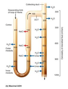
The nephron loop (Loop of Henle) establishes a solute concentration gradient in the medulla, which is essential for water reabsorption and urine concentration. The descending limb is permeable to water, while the ascending limb is permeable to NaCl.
ADH (Antidiuretic Hormone): Increases water reabsorption by inserting aquaporins in the DCT and collecting duct, allowing urine to become highly concentrated.
Without ADH: No aquaporins are present, resulting in a large volume of dilute urine.
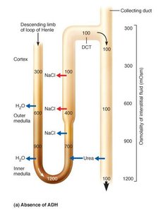
Reabsorption of Glucose
Glucose is reabsorbed in the nephron by carrier proteins. Under normal conditions, all glucose is reabsorbed and should not appear in urine. If the number of glucose carriers is insufficient, glucose may be excreted in urine.
Carrier-mediated transport: Glucose reabsorption depends on the number of carrier proteins.
Clinical relevance: Presence of glucose in urine may indicate diabetes mellitus.
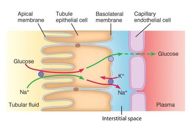
Effects of Hormones on Renal Function
Two key hormones regulate renal function and urine composition:
Antidiuretic Hormone (ADH): Promotes water reabsorption, resulting in concentrated urine.
Aldosterone: Increases sodium reabsorption and potassium secretion, affecting urine volume and electrolyte balance.
Both hormones: Increase blood pressure by increasing blood volume.
Aldosterone | ADH | Urine Volume (mL) | Urine Concentration (mOsm) | Potassium in Urine (mM) |
|---|---|---|---|---|
Absent | Absent | 201.00 | 100 | 6.25 |
Present | Absent | 180.90 | 100 | 10.42 |
Absent | Present | 16.86 | 1200 | 62.37 |
Present | Present | 12.67 | 1200 | 65.37 |
Key Points:
Aldosterone decreases urine volume by increasing sodium reabsorption.
ADH increases urine concentration by promoting water reabsorption.
Both hormones affect potassium concentration in urine due to changes in sodium and water handling.
Summary Table: Solute Gradient and Urine Concentration
Solute Concentration Gradient (mOsm) | Urine Concentration (mOsm) | Urine Volume (mL) |
|---|---|---|
300 | 300 | 80.57 |
600 | 600 | 40.28 |
900 | 900 | 26.86 |
1200 | 1200 | 16.86 |
Formula for GFR:
Kf: Filtration coefficient
PGC: Glomerular capillary hydrostatic pressure
PBS: Bowman's space hydrostatic pressure
πGC: Glomerular capillary oncotic pressure
Additional info: The notes expand on the physiological mechanisms underlying renal filtration, solute gradients, and hormonal regulation, providing context for laboratory and clinical applications.
