 Indietro
Study Guide: Sense Organs and Special Senses in Anatomy & Physiology
Guida di studio - Note intelligenti
 Appunti personalizzati basati sui tuoi materiali, ampliati con definizioni chiave, esempi e contesto.
Sense Organs and Sensory Receptors
Introduction to Sensory Receptors
Sensory receptors are specialized cells or nerve endings that detect changes (stimuli) in the internal or external environment and convert them into nerve signals interpretable by the nervous system. These signals allow the body to perceive touch, pressure, pain, temperature, stretch, light, sound, and chemicals.
Definition: Sensory receptors transduce environmental stimuli into electrical signals.
Examples: Touch, pain, temperature, light, sound, taste, and smell.
Classification of Sensory Receptors
Sensory receptors are classified by stimulus modality, origin of stimulus, and distribution in the body.
By Stimulus Modality:
Thermoreceptors: Detect temperature changes.
Photoreceptors: Respond to light (e.g., retina).
Nociceptors: Detect pain from tissue injury.
Chemoreceptors: Respond to chemicals (taste, smell, pH).
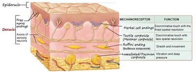
Mechanoreceptors: Respond to physical deformation (touch, pressure, vibration, stretch).
By Origin of Stimulus:
Exteroceptors: External environment (vision, hearing, taste, smell, cutaneous sensations).
Interoceptors: Internal environment (stretch, pressure, visceral pain, nausea).
Proprioceptors: Body position and movement (muscles, tendons, joints).
By Distribution:
General (Somatosensory) Senses: Widely distributed (skin, muscles, tendons, joints, viscera).
Special Senses: Limited to head, involve complex sense organs (vision, hearing, equilibrium, taste, smell).
Structural Organization of Somatosensory Receptors
Unencapsulated and Encapsulated Receptors
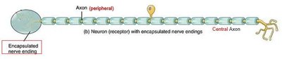
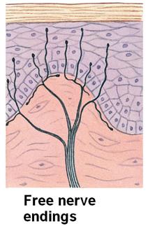
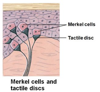
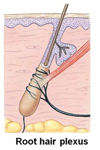
Somatosensory receptors are peripheral endings of unipolar sensory neurons. They may be unencapsulated (no connective tissue wrapping) or encapsulated (wrapped in glial cells or connective tissue).
Unencapsulated: Free nerve endings, tactile (Merkel) discs, hair receptors (root hair plexuses).
Encapsulated: Mechanoreceptors such as tactile corpuscles, end bulbs, bulbous corpuscles, lamellar corpuscles.


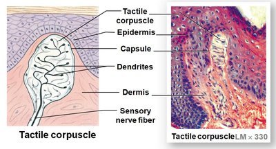
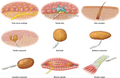
Processing of Sensory Information
Sequence from Stimulus to Perception
Sensory information is processed through a series of steps: stimulus, detection, transduction, transmission, and perception.
Stimulus: Activates a sensory receptor.
Detection: Receptor responds to the stimulus.
Transduction: Stimulus energy is converted into an electrical signal.
Transmission: Action potential travels to the CNS.
Perception: Brain interprets the sensory information.
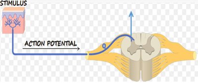
Somatosensory Projection Pathways
Spinal Tracts and Pathways
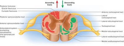
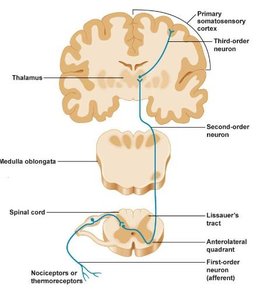
Spinal tracts are bundles of axons in the white matter of the spinal cord. Ascending tracts carry sensory information, while descending tracts carry motor signals. Sensory signals travel across three neurons from origin to destination in the brain.
First-order neuron: Detects stimulus, transmits signal to spinal cord or brainstem.
Second-order neuron: Continues to thalamus, often decussates (crosses over).
Third-order neuron: Carries signal to the primary somatosensory cortex.
Special Senses: Taste (Gustation)
Anatomical Structures Involved in Taste
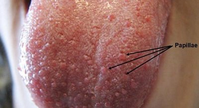
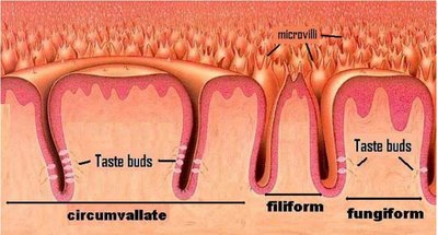
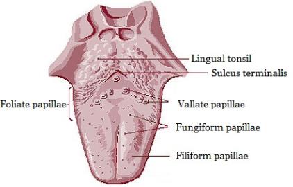
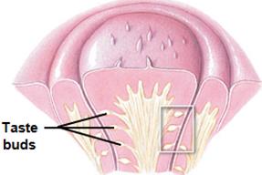
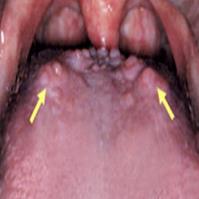
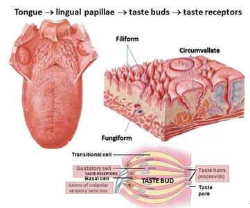
The sense of taste involves the tongue, lingual papillae, taste buds, and taste receptor cells. Taste buds are located on specific papillae and contain gustatory cells.
Tongue: Contains lingual papillae.
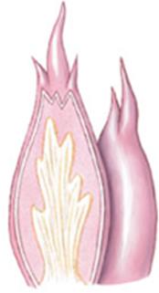
Lingual papillae: Four types—fungiform, circumvallate, filiform, foliate.
Taste buds: Contain gustatory receptor cells.
Taste receptor cells: Chemoreceptors activated by tastants dissolved in saliva.
Types of Cells in Taste Buds
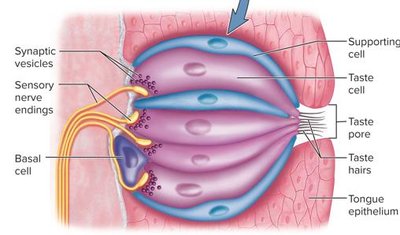
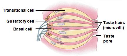
Taste buds consist of basal cells (stem cells), supporting cells, and gustatory cells. Gustatory microvilli (taste hairs) are the site of taste detection.
Basal cells: Regenerate into gustatory cells.
Supporting cells: Surround and support gustatory cells.
Gustatory cells: Have taste hairs (microvilli) with taste receptors.
Taste hairs: Detect tastants dissolved in saliva.
Classes of Taste Sensations
There are five primary taste sensations: sour, salty, bitter, sweet, and umami. Taste is closely linked to smell, which contributes significantly to flavor perception.
Sour
Salty
Bitter
Sweet
Umami
Special Senses: Smell (Olfaction)
Olfactory Epithelium and Receptors
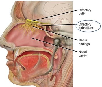
The olfactory epithelium is located in the roof of the nasal cavity and contains cells specialized for detecting odorants in the air.
Cell types: Basal (stem) cells, supporting cells, olfactory neurons (bipolar).
Olfactory receptors: Chemoreceptors located on olfactory cilia, activated by odorant molecules dissolved in mucus.
Olfactory Cilia and Pathway
Olfactory cilia are hair-like projections on the dendrites of olfactory neurons, where odorant detection occurs. The olfactory nerve (CN I) carries signals to the olfactory bulb and tract. Olfactory information does not synapse in the thalamus before reaching the cerebral cortex.
Special Senses: Hearing and Equilibrium
Anatomy of the Ear
The ear consists of three principal regions: outer ear, middle ear, and inner ear. Each region has specialized structures for hearing and balance.
Outer ear: Auricle (pinna), auditory canal; directs sound waves to tympanic membrane.
Middle ear: Tympanic membrane, auditory ossicles (malleus, incus, stapes), auditory (Eustachian) tube; amplifies and transmits sound.
Inner ear: Bony and membranous labyrinth, vestibular complex (balance), cochlea (hearing).
Special Senses: Vision
Accessory and Internal Structures of the Eye
The eye consists of accessory structures (protection, lubrication, support) and internal structures (vision).
Accessory structures: Eyebrows, eyelashes, eyelids, conjunctiva, lacrimal apparatus, extrinsic eye muscles.
Internal structures: Lens, two cavities, three tunics (fibrous, vascular, neural).
Eye Tunics (Layers)
Fibrous tunic: Sclera (white of the eye), cornea (transparent, focuses light).
Vascular tunic: Choroid (blood supply), ciliary body (lens shape, aqueous humor), iris (light regulation, eye color).
Neural tunic (retina): Photoreceptors (rods and cones), bipolar cells, ganglion cells; optic nerve formation; macula lutea (central vision), optic disc (blind spot).
Visual Pathway
Light passes through cornea, aqueous humor, lens, vitreous humor, and retina. Action potentials travel via the optic nerve, optic chiasm, and optic tract to the visual cortex.
Cornea: Refraction
Aqueous humor
Lens: Refraction
Vitreous humor
Retina: Photoreceptors, bipolar cells, ganglion cells
Optic nerve: Axons of ganglion cells
Optic chiasm: Partial crossover of fibers
Visual cortex: Perception and interpretation
Mechanoreceptor | Function |
|---|---|
Merkel cell ending | Discriminative touch with the finest spatial resolution |
Tactile corpuscle (Meissner corpuscle) | Discriminative touch with less spatial resolution |
Ruffini ending (Bulbous corpuscle) | Stretch and movement |
Lamellated/Pacinian corpuscle | Vibration and deep pressure |
Additional info: Academic context was added to clarify the structural and functional relationships of sensory receptors, pathways, and special senses, as well as to ensure completeness and self-contained explanations for exam preparation.
