 Indietro
The Appendicular Skeleton: Structure and Function
Guida di studio - Note intelligenti
 Appunti personalizzati basati sui tuoi materiali, ampliati con definizioni chiave, esempi e contesto.
The Appendicular Skeleton
Overview
The appendicular skeleton consists of 126 bones that form the limbs and their girdles, enabling movement and manipulation of the environment. It is divided into the pectoral girdle (shoulder), upper limb, pelvic girdle (hip), and lower limb.
Pectoral girdle attaches the upper limbs to the trunk.
Pelvic girdle attaches the lower limbs to the trunk.
Each limb has three major segments connected by movable joints.
Pectoral Girdle
Structure and Function
The pectoral girdle, also known as the shoulder girdle, is composed of the clavicle and scapula. It attaches the upper limb to the axial skeleton and provides attachment sites for muscles that move the upper limbs.
Clavicle: S-shaped bone, only bone of the shoulder girdle/upper limb that directly attaches to the axial skeleton.
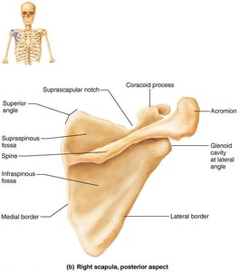
Scapula: Flat, triangular bone located on the posterior surface of the rib cage.
Shoulder girdles and associated muscles form the shoulders.
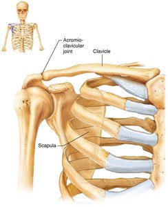
Clavicle
The clavicle is an S-shaped bone with a cone-shaped sternal end that articulates with the sternum and a flattened acromial end that articulates with the scapula.
Anchors muscles and braces the scapula laterally.
Transmits compressive forces from the upper limb to the axial skeleton.
Fractures typically occur anteriorly; posterior fractures may endanger the subclavian artery.
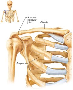
Scapula
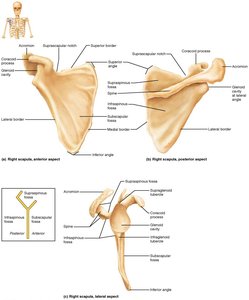
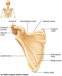
The scapula is a flat, triangular bone with three borders and three angles. It contains several large fossae named according to their location. The glenoid fossa is shallow, allowing for movement but also instability.
Located between ribs 2 and 7 on the posterior rib cage.
Provides attachment points for muscles and articulates with the humerus.
Upper Limb
Structure
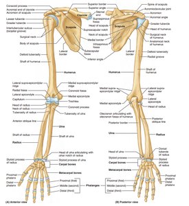
Each upper limb consists of 30 bones, forming the arm, forearm, and hand.
Arm: Humerus (largest, longest bone of the upper limb).
Forearm: Radius (lateral) and ulna (medial).
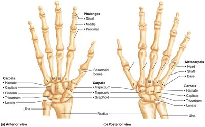
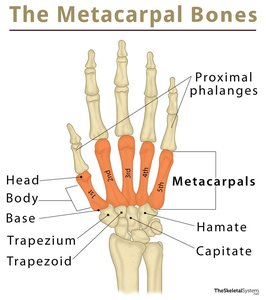
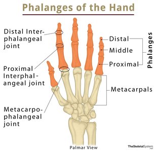
Hand: 8 carpal bones (wrist), 5 metacarpal bones (palm), 14 phalanges (fingers).
Humerus
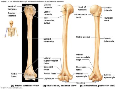
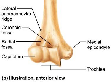
The humerus is the largest and longest bone of the upper limb. Its head articulates with the glenoid cavity of the scapula, and its condyles (trochlea and capitulum) articulate with the radius and ulna.
Provides attachment sites for muscles.
Forms the elbow joint with the ulna and radius.
Forearm: Ulna and Radius
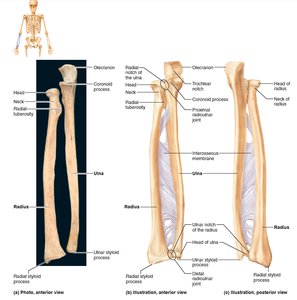
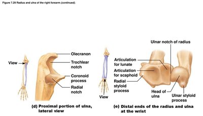
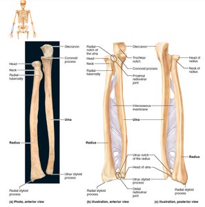
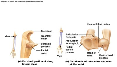
The forearm consists of the ulna (medial) and radius (lateral), connected by an interosseous membrane.
Ulna: Contributes more to the elbow joint; forms the major portion of the elbow joint with the humerus.
Radius: Contributes more to the wrist joint; hand moves with the radius.
Wrist and Hand
The wrist (carpus) consists of 8 carpal bones arranged in two rows. The hand includes 5 metacarpal bones and 14 phalanges.
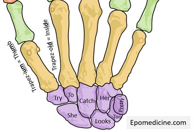
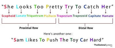
Carpal bones: Proximal row (scaphoid, lunate, triquetrum, pisiform); distal row (trapezium, trapezoid, capitate, hamate).
Metacarpals: Numbered I to V from thumb to little finger.
Phalanges: Fingers numbered I to V; thumb (pollex) has 2 bones, other fingers have 3 bones each.
Pelvic Girdle
Structure and Function
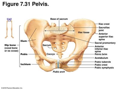
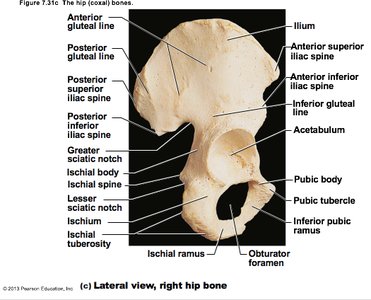
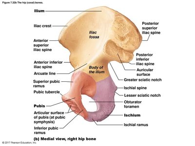
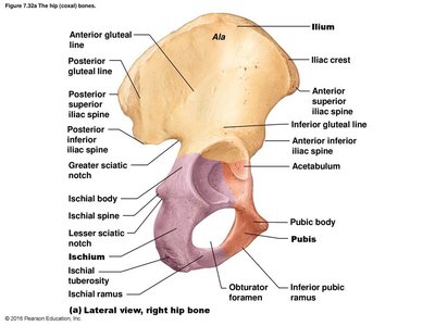
The pelvic girdle consists of two hip bones (coxal bones) and the sacrum. It attaches the lower limbs to the axial skeleton, transmits weight, and supports pelvic organs.
Three fused bones form each coxal bone: ilium, ischium, and pubis.
Less mobility but more stability than the shoulder joint.
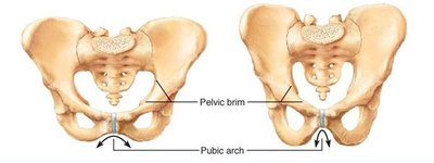
Comparison of Male and Female Pelvis
The female pelvis is adapted for childbearing, with a wide, shallow, and round cavity. The male pelvis is adapted for supporting a heavier build and stronger muscles, with a narrow and deep cavity.
Female pelvis: Greater capacity, wider pelvic brim, broader pubic arch.
Male pelvis: Heavier, narrower pelvic brim, sharper pubic arch.
Lower Limb
Structure
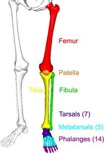
The lower limb bears the weight of the body and is stronger and thicker than the upper limb. It consists of three segments: thigh, leg, and foot.
Thigh: Femur (largest and strongest bone in the body), patella (sesamoid bone in quadriceps tendon).
Leg: Tibia (medial, weight-bearing), fibula (lateral, limited weight-bearing).
Foot: Tarsals (7 bones), metatarsals (5 bones), phalanges (14 bones).
Thigh: Femur and Patella
Femur: Articulates proximally with the acetabulum of the hip and distally with the tibia and patella.
Patella: Protects the knee joint and improves leverage of the thigh muscles.
Leg: Tibia and Fibula
Tibia: Receives weight from the femur and transmits it to the foot.
Fibula: Does not articulate with the femur; provides lateral stability.
Tibia and fibula are connected by an interosseous membrane.
Foot: Tarsals, Metatarsals, and Phalanges
Tarsals: 7 bones, including calcaneus (heel), talus (articulates with tibia), navicular, cuboid, and three cuneiforms.
Metatarsals: 5 bones, numbered I to V from hallux (big toe) to little toe.
Phalanges: Digit I (hallux) has 2 bones; digits II to V have 3 bones each.
Arches of the Foot
The foot has three arches maintained by interlocking bones, ligaments, and tendons, allowing it to bear weight and act as a lever.
Lateral longitudinal arch
Medial longitudinal arch
Transverse arch
Developmental Aspects of the Skeleton
Babies and Children
Skull has fontanelles; head is large relative to body.
Full cranium size by 8-9 years old; face grows between 6-13 years old.
Convex curvatures of the spine present at birth; secondary curvatures develop with growth.
Old Age
Intervertebral discs thin, less hydrated, and less elastic; risk of disc herniation increases.
Height loss common by age 55; costal cartilages ossify.
Rigid thorax causes shallow breathing and less efficient gas exchange.
All bones lose mass, increasing fracture risk.
Summary Table: Bones of the Appendicular Skeleton
Region | Main Bones | Key Features |
|---|---|---|
Pectoral Girdle | Clavicle, Scapula | Attaches upper limb to trunk |
Upper Limb | Humerus, Radius, Ulna, Carpals, Metacarpals, Phalanges | Movement, manipulation |
Pelvic Girdle | Ilium, Ischium, Pubis, Sacrum | Attaches lower limb, supports organs |
Lower Limb | Femur, Patella, Tibia, Fibula, Tarsals, Metatarsals, Phalanges | Weight bearing, locomotion |
Key Terms and Definitions
Girdle: Structure that attaches limbs to the axial skeleton.
Fossa: Shallow depression or hollow in a bone.
Condyle: Rounded articular surface.
Sesamoid bone: Bone embedded within a tendon.
Formulas and Equations
Bone count in appendicular skeleton:
Examples and Applications
Fracture of the clavicle can compromise the subclavian artery if displaced posteriorly.
Female pelvis adaptations facilitate childbirth.
Arches of the foot allow efficient weight distribution and movement.
Additional info:
Mnemonic devices for carpal bones aid memorization for anatomy students.
Developmental changes in the skeleton are important for understanding pediatric and geriatric anatomy.
