Indietro
The Axial Skeleton: Structure and Function
Guida di studio - Note intelligenti
 Appunti personalizzati basati sui tuoi materiali, ampliati con definizioni chiave, esempi e contesto.
The Axial Skeleton
Overview of the Human Skeleton
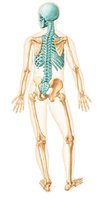
The human skeleton is a framework of bones, cartilage, joints, and ligaments that supports and protects the body. It is divided into two main divisions: the axial skeleton and the appendicular skeleton. The axial skeleton consists of 80 bones, including the skull, vertebral column, and thoracic cage, and serves as the central axis of the body.
Axial skeleton: Skull, vertebral column, thoracic cage
Appendicular skeleton: Limbs and girdles (see Chapter 8)
The Skull
Structure and Function
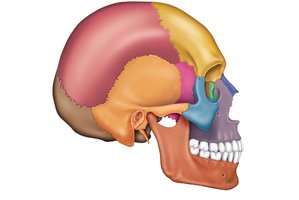
The skull is the most complex bony structure in the body, formed by cranial and facial bones. The cranium encloses and protects the brain and provides attachment sites for head and neck muscles. Facial bones form the framework of the face, house sense organs, provide openings for air and food, hold teeth, and anchor facial muscles.
Cranial bones: Enclose the brain
Facial bones: Form the face and support sensory structures
Sutures of the Skull
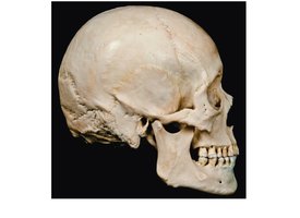
Most skull bones are flat and joined by immovable joints called sutures, which have an irregular, saw-toothed appearance. Major sutures include the coronal, squamous, sagittal, and lambdoid sutures.
Skull Geography
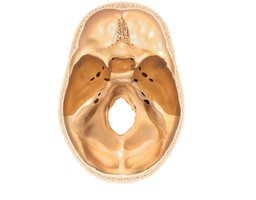
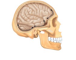
The skull is divided into the cranial vault (calvaria or skull cap) and the cranial base. Internally, the cranial base is divided into anterior, middle, and posterior cranial fossae, which house different regions of the brain. The skull also contains smaller cavities such as the orbits, nasal cavity, and paranasal sinuses, and approximately 85 named openings for nerves and blood vessels.
Cranial Bones
Major Cranial Bones
The cranium is formed from eight large bones: paired parietal and temporal bones, and unpaired frontal, occipital, sphenoid, and ethmoid bones. These bones are joined by sutures and provide protection and structural support for the brain.
Parietal Bones and Sutures
Parietal bones form the superior and lateral aspects of the skull. The four major sutures associated with the parietal bones are:
Coronal suture: Parietal bones meet the frontal bone
Squamous suture: Parietal bones meet temporal bones
Sagittal suture: Right and left parietal bones meet
Lambdoid suture: Parietal bones meet the occipital bone
Occipital Bone
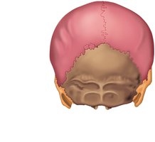
The occipital bone forms the posterior part of the cranium and cranial base. It articulates with the temporal and parietal bones and forms the posterior cranial fossa. Key features include the foramen magnum, occipital condyles (for head nodding), and external occipital protuberance (ligament attachment).
Temporal Bones
Temporal bones lie inferior to the parietal bones and form the inferolateral portion of the skull. They consist of squamous, tympanic, and petrous parts, and contain important foramina for nerves and blood vessels. The mastoid process serves as a muscle attachment site, and the petrous part houses the middle and inner ear.
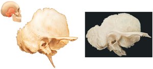
Sphenoid Bone
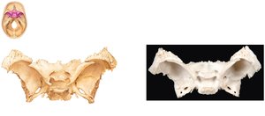
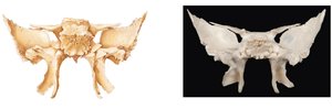
The sphenoid bone spans the width of the cranial floor and is considered the "keystone" of the cranium. It consists of a body, greater and lesser wings, and pterygoid processes. The sella turcica houses the pituitary gland. The sphenoid contains several important foramina for cranial nerves and blood vessels.
Ethmoid Bone
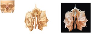
The ethmoid bone lies between the nasal and sphenoid bones and forms much of the medial bony region between the nasal cavity and orbits. Key features include the cribriform plate (with foramina for olfactory nerves), crista galli (membrane attachment), perpendicular plate (nasal septum), and ethmoidal labyrinth (air cells).
Facial Bones
Major Facial Bones
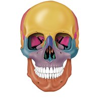
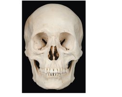
The facial skeleton consists of 14 bones: the unpaired mandible and vomer, and paired maxillae, zygomatic, nasal, lacrimal, palatine, and inferior nasal conchae. These bones form the structure of the face, house the teeth, and contribute to the orbits and nasal cavity.
Mandible
The mandible is the largest and strongest facial bone and the only movable bone of the skull. It consists of a horizontal body and two upright rami. Major landmarks include the mandibular foramen, alveolar process, mental foramen, condylar process, and ramus.
Maxillary Bones
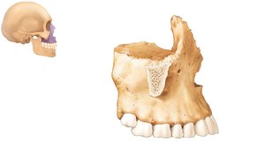
The maxillae articulate with all other facial bones except the mandible and contain the largest paranasal sinuses. They form the upper jaw and part of the hard palate, and are considered the "keystone" bones of the face.
Other Facial Bones
Zygomatic bones: Form the lateral wall of the orbits
Nasal bones: Form the bridge of the nose
Lacrimal bones: Located in the medial orbital walls
Palatine bones: Complete the posterior part of the hard palate
Vomer: Forms the inferior part of the nasal septum
Inferior nasal conchae: Project medially from the lateral walls of the nasal cavity
Special Parts of the Skull
Nasal Cavity and Paranasal Sinuses
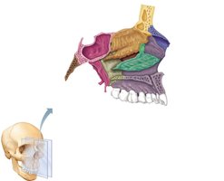
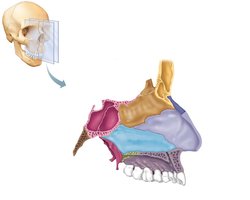
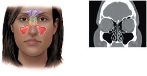
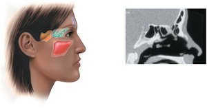
The nasal cavity is formed by the ethmoid, maxillae, and palatine bones. Paranasal sinuses are air-filled spaces within the frontal, ethmoid, sphenoid, and maxillary bones. They are lined with mucous membrane, help warm and moisten air, lighten the skull, and enhance voice resonance.
Orbits
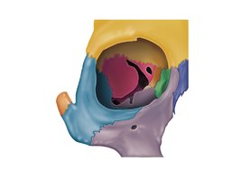
The orbits are bony cavities that house the eyeballs and are formed by seven bones: frontal, sphenoid, zygomatic, maxilla, palatine, lacrimal, and ethmoid.
Hyoid Bone
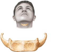
The hyoid bone lies inferior to the mandible and is unique in that it does not articulate directly with any other bone. It serves as a movable base for the tongue and an attachment site for muscles associated with swallowing and speech.
The Vertebral Column
Structure and Function
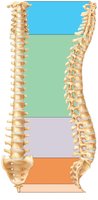
The vertebral column is composed of 26 bones in adults and serves to transmit the weight of the trunk to the lower limbs, protect the spinal cord, and provide attachment sites for muscles. It is divided into five regions: cervical, thoracic, lumbar, sacral, and coccygeal.
Regions and Curvatures
Cervical (7 vertebrae): Neck region, concave posteriorly
Thoracic (12 vertebrae): Upper back, convex posteriorly
Lumbar (5 vertebrae): Lower back, concave posteriorly
Sacrum: 5 fused vertebrae, convex posteriorly
Coccyx: 3–5 fused vertebrae
Ligaments and Intervertebral Discs
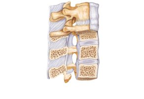
Major ligaments support the vertebral column, including the anterior and posterior longitudinal ligaments and the ligamentum flavum. Intervertebral discs act as shock absorbers and are composed of a gelatinous nucleus pulposus and a strong anulus fibrosus.
General Structure of Vertebrae
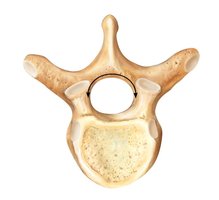
All vertebrae share common features: a body, vertebral arch, vertebral foramen, spinous and transverse processes, and superior/inferior articular processes. The structure varies by region to accommodate different functions and movements.
Cervical Vertebrae
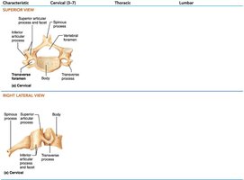
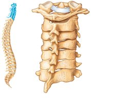
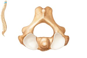
Cervical vertebrae are the smallest and lightest. They have a wide body, short bifid spinous processes, large triangular vertebral foramen, and transverse foramina. The atlas (C1) supports the skull and allows nodding, while the axis (C2) has a dens for rotation.
Thoracic Vertebrae
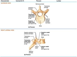
Thoracic vertebrae articulate with ribs, have heart-shaped bodies, long spinous processes, and circular vertebral foramina. Their transverse processes articulate with rib tubercles, and their articular facets allow rotation but limit flexion and extension.
Lumbar Vertebrae
Lumbar vertebrae have thick, robust bodies, short and flat spinous processes, and triangular vertebral foramina. Their articular facets are oriented medially, allowing flexion and extension but preventing rotation.
Sacrum and Coccyx
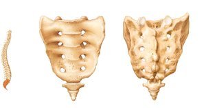
The sacrum is formed from five fused vertebrae and shapes the posterior wall of the pelvis. It contains sacral foramina for nerve passage, a median sacral crest, and the sacral canal. The coccyx, or tailbone, is formed from 3–5 fused vertebrae and provides slight support to pelvic organs.
The Thoracic Cage
Structure and Function
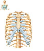
The thoracic cage forms the bony framework of the chest, consisting of thoracic vertebrae, ribs, sternum, and costal cartilage. It protects thoracic organs, supports the shoulder girdle and upper limbs, and provides muscle attachment sites.
Sternum
The sternum is composed of the manubrium, body, and xiphoid process. Key landmarks include the jugular notch, sternal angle, and xiphisternal joint. The sternum articulates with the clavicles and costal cartilages of the ribs.
Ribs
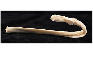
There are 12 pairs of ribs. True ribs (1–7) attach directly to the sternum via costal cartilage. False ribs (8–12) do not attach directly; ribs 11–12 are floating ribs. Ribs articulate posteriorly with the vertebral column.
The Axial Skeleton Throughout Life
Development and Aging
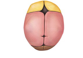
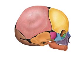
Membrane bones of the skull begin to ossify in the second month of development. Fontanelles are unossified remnants that allow for skull compression during birth and brain growth. With aging, intervertebral discs lose water content, the thorax becomes more rigid, and bone mass decreases.