 Indietro
The Cell: Structure, Function, and Membrane Transport
Guida di studio - Note intelligenti
 Appunti personalizzati basati sui tuoi materiali, ampliati con definizioni chiave, esempi e contesto.
The Cell
General Structure of Cells
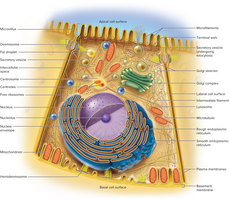
The cell is the basic structural and functional unit of all living organisms. Human cells exhibit a wide variety of shapes, sizes, and specialized functions, with approximately 200 different types present in the body.
Plasma membrane: The boundary that separates the cell from its environment.
Cytoplasm: The internal fluid containing organelles and cytoskeleton.
Nucleus: The control center containing DNA.
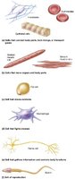
Examples of cell types: Epithelial cells, muscle cells, nerve cells, and more, each adapted for specific roles.
Cell Components
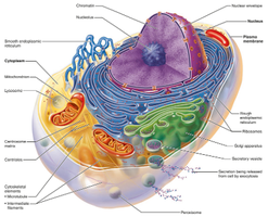
Each cell contains specialized structures called organelles, which perform distinct functions necessary for cell survival and activity.
Nucleus: Contains genetic material and directs cellular activities.
Endoplasmic reticulum (ER): Synthesizes proteins (rough ER) and lipids (smooth ER).
Golgi apparatus: Modifies, sorts, and packages proteins and lipids.
Mitochondria: Site of ATP (energy) production.
Lysosomes: Contain digestive enzymes for breaking down waste.
Peroxisomes: Detoxify harmful substances.
Cytoskeleton: Provides structural support and facilitates movement.
The Cell Surface
Plasma Membrane Structure
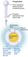
The plasma membrane is a dynamic, selectively permeable barrier composed mainly of a phospholipid bilayer, cholesterol, and glycolipids. It separates the intracellular fluid (ICF) from the extracellular fluid (ECF).
Phospholipids: Amphipathic molecules with hydrophilic heads and hydrophobic tails.
Cholesterol: Stabilizes membrane fluidity.
Glycolipids: Contribute to cell recognition and signaling.
Membrane Proteins
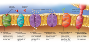
Membrane proteins are crucial for the diverse functions of the plasma membrane. They can be classified as peripheral (attached to the membrane surface) or integral (span the membrane).
Channel proteins: Form pores for selective passage of ions and molecules.
Gated channels: Open or close in response to stimuli.
Receptors: Bind signaling molecules (e.g., hormones) to initiate cellular responses.
Enzymes: Catalyze reactions at the membrane surface.
Cell identity markers: Distinguish self from foreign cells.
Cell adhesion molecules (CAMs): Mediate cell-to-cell attachment.
Cell Surface Extensions
Cells may possess surface extensions that increase surface area or aid in movement:
Microvilli: Increase surface area for absorption.
Cilia: Move substances across the cell surface.
Flagella: Propel cells (e.g., sperm).
Pseudopods: Temporary projections for movement or engulfing particles.
Cell Junctions
Cell junctions are protein complexes that connect cells to each other and to the extracellular matrix, enabling communication, structural integrity, and coordinated function.
Tight junctions: Seal adjacent cells to prevent leakage of extracellular fluid.
Desmosomes: Anchor cells together, providing mechanical strength.
Gap junctions: Allow direct passage of ions and small molecules between cells for rapid communication.
Membrane Transport
Overview of Membrane Transport
Cells regulate the movement of substances across their membranes through various transport mechanisms, which are essential for maintaining homeostasis.
Passive transport: Does not require energy (ATP).
Active transport: Requires energy input (ATP).
Filtration
Filtration is the process by which physical pressure forces fluid and small solutes through a selectively permeable membrane, while larger particles are retained.
Simple Diffusion
Simple diffusion is the movement of particles from an area of higher concentration to an area of lower concentration, down their concentration gradient. It does not require energy and is used by small, non-polar molecules such as oxygen and carbon dioxide.
Osmosis
Osmosis is the net movement of water across a selectively permeable membrane from an area of low solute concentration to an area of high solute concentration. It is vital for maintaining cell volume and pressure.
Tonicity: The ability of a solution to affect cell volume and pressure by osmosis.
Isotonic: Equal solute concentration inside and outside the cell; no net water movement.
Hypertonic: Higher solute concentration outside the cell; cell loses water and shrivels.
Hypotonic: Lower solute concentration outside the cell; cell gains water and may burst.
Carrier-Mediated Transport
Carrier proteins facilitate the movement of specific solutes across the membrane. There are two main types:
Facilitated diffusion: Solute moves down its concentration gradient with the help of a carrier protein; no energy required.
Active transport: Solute moves against its concentration gradient using a carrier protein and ATP.
Vesicular Transport
Vesicular transport involves the movement of large particles or fluids via vesicles and requires ATP.
Endocytosis: Bringing substances into the cell.
Exocytosis: Expelling substances from the cell.
Phagocytosis: "Cell eating"; engulfing large particles.
Pinocytosis: "Cell drinking"; engulfing extracellular fluid.
Receptor-mediated endocytosis: Selective uptake of specific molecules via receptor binding.
The Cell Interior
Organelles
Organelles are specialized structures within the cell that perform distinct metabolic functions, maintaining cellular order and efficiency.
Cytoskeleton
The cytoskeleton is a network of protein filaments that provides structural support, determines cell shape, and facilitates movement.
Microfilaments: Thin filaments involved in cell movement and shape.
Intermediate filaments: Provide mechanical strength.
Microtubules: Hollow tubes that guide organelle movement and form the mitotic spindle.
Nucleus
The nucleus is the largest organelle, surrounded by a double membrane (nuclear envelope). It contains chromosomes and the nucleolus, where ribosome subunits are assembled.
Endoplasmic Reticulum (ER)
The ER is a network of membranes with two regions:
Rough ER: Studded with ribosomes; synthesizes proteins for secretion or membrane insertion.
Smooth ER: Lacks ribosomes; synthesizes lipids, detoxifies chemicals, and stores calcium.
Ribosomes
Ribosomes are composed of RNA and proteins. They read mRNA and assemble amino acids into proteins. Free ribosomes produce proteins for the cytosol; bound ribosomes (on RER) produce proteins for export.
Golgi Complex
The Golgi apparatus modifies, sorts, and packages proteins and lipids for delivery to various destinations. It forms vesicles that may become lysosomes, secretory vesicles, or fuse with the plasma membrane.
Lysosomes
Lysosomes are membrane-bound sacs containing digestive enzymes. They break down macromolecules, cellular debris, and play a role in apoptosis (programmed cell death).
Peroxisomes
Peroxisomes contain enzymes that detoxify harmful substances and neutralize free radicals. They break down fatty acids and convert hydrogen peroxide to water.
Mitochondria
Mitochondria are the "powerhouses" of the cell, generating ATP through aerobic respiration. They have a double membrane, with the inner membrane folded into cristae, and contain their own DNA.
Centrioles
Centrioles are cylindrical structures that organize microtubules during cell division, forming the mitotic spindle. A pair of centrioles is found in the centrosome near the nucleus.
The Cell Life Cycle
Phases of the Cell Cycle
The cell cycle consists of interphase (G1, S, G2) and the mitotic phase (mitosis and cytokinesis).
G1 phase: Cell grows and performs normal functions; centrioles begin to replicate.
S phase: DNA is replicated in preparation for cell division.
G2 phase: Cell prepares for division; centrioles finish replicating.
Mitosis: Division of the nucleus and its DNA into two identical sets.
Cytokinesis: Division of the cytoplasm, resulting in two daughter cells.
Mitosis Stages
Prophase: Chromosomes condense, nuclear envelope breaks down, spindle fibers form.
Metaphase: Chromosomes align at the cell's equator.
Anaphase: Sister chromatids separate and move to opposite poles.
Telophase: Chromatids decondense, nuclear envelopes reform, cytokinesis begins.
Clinical Connection: Tay-Sachs Disease
Tay-Sachs disease is a genetic disorder caused by a deficiency of a lysosomal enzyme needed to break down a specific glycolipid in nerve cells. Accumulation of this glycolipid leads to neurodegeneration, loss of developmental milestones, and early death.
Key Terms and Concepts
Amphipathic: Molecules with both hydrophilic and hydrophobic regions (e.g., phospholipids).
ATP: Adenosine triphosphate, the main energy currency of the cell.
Homeostasis: Maintenance of a stable internal environment.
