 Indietro
The Integumentary System: Accessory Organs and Clinical Applications
Guida di studio - Note intelligenti
 Appunti personalizzati basati sui tuoi materiali, ampliati con definizioni chiave, esempi e contesto.
Integumentary System: Accessory Organs
Overview of Accessory Organs
The accessory organs of the skin include hair, nails, and cutaneous glands. These structures arise as modifications or invaginations of the epidermis during embryonic development, although their mature forms often extend into the dermis. Understanding their structure and function is essential for comprehending the protective and regulatory roles of the skin.
Cutaneous Glands
The skin contains five main types of exocrine glands, all derived from epithelial tissue:
Merocrine (Eccrine) Sweat Glands
Apocrine Sweat Glands
Sebaceous Glands
Ceruminous Glands
Mammary Glands
Each gland type releases its products by distinct mechanisms and is located in specific regions of the skin.
Sweat Glands
Sweat glands, also known as sudoriferous glands, are distributed across nearly all skin surfaces except the nipples and parts of the external genitalia. There are approximately 3 million sweat glands per person, classified into four types:
Eccrine (Merocrine)
Apocrine
Ceruminous (modified)
Mammary (modified)
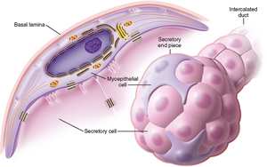
These glands contain myoepithelial cells that contract to force secretion into ducts.
Merocrine (Eccrine) Sweat Glands
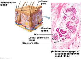
Merocrine glands are the most numerous, smaller, and more superficial than apocrine glands. They are abundant on the palms, soles, and forehead. Their acini are composed of simple cuboidal epithelial tissue, and their ducts open directly onto the skin surface. The primary function is thermoregulation, regulated by the sympathetic nervous system. Their secretion is mostly water (99%), with salts, vitamin C, antibodies, dermcidin, and metabolic wastes.
Location: Reticular layer of dermis
Secretion: Exocytosis
Function: Thermoregulation
Apocrine Sweat Glands
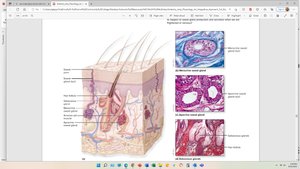
Apocrine glands are mostly found in the axillae, areolae, pubic, and perineal regions. They secrete a viscous, milky sweat containing fatty substances and proteins, which bacteria break down to produce body odor. These glands are larger and deeper than eccrine glands, and their ducts empty into hair follicles. They begin functioning at puberty and are considered vestigial scent glands.
Location: Reticular layer of dermis, near hair follicles
Secretion: Exocytosis
Function: Scent, body odor
Sebaceous Glands
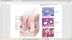
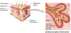
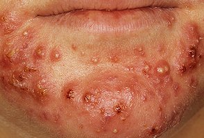
Sebaceous glands produce sebum, an oily secretion that keeps skin and hair from becoming dry, brittle, or cracked. These flask-shaped glands open into hair follicles and are most concentrated on the face, scalp, upper chest, and back. They are holocrine glands, releasing their products by rupture of the plasma membrane.
Location: Associated with hair follicles
Secretion: Holocrine (cell rupture)
Function: Lubrication, protection
Ceruminous Glands
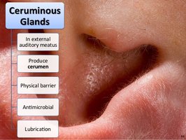
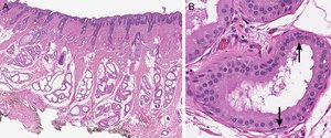
Ceruminous glands are modified apocrine sweat glands located in the external auditory canal. Their secretions combine with sebaceous secretions and desquamated keratinocytes to form cerumen (earwax). Cerumen acts as a water-repellent barrier, protects against physical damage, inhibits microbial growth, and prevents insect invasion.
Location: External auditory canal
Secretion: Cerumen (earwax)
Function: Protection, antimicrobial
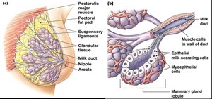
Mammary Glands
Mammary glands are present in both sexes but are responsible for milk production (lactation) in females. Glandular tissue develops after puberty in response to hormones. These glands use both apocrine (lipid droplets) and merocrine (proteins and sugars) secretion mechanisms.
Location: Hypodermis, breast tissue
Secretion: Milk (lipids, proteins, sugars)
Function: Nutrition for offspring
Hair
Structure and Function of Hair
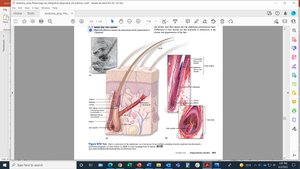
The hair follicle is an invagination of the epidermis into the dermis. At its base is the hair bulb, which contains the hair matrix—the region of active cell division responsible for hair growth. The hair bulb is supplied by a vascular connective tissue dermal papilla, providing nutrients and growth signals. As matrix cells divide, they are pushed upward, become keratinized, and form the hair shaft. Melanocytes in the matrix produce melanin, giving hair its color.
Functions: Insulation, protection, sensory warning, social signaling
Hair bulb: Contains viable cells and matrix
Hair shaft: Dead, keratinized cells
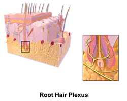
Root hair plexus: Sensory nerve endings
Hair Matrix and Papilla
The hair matrix is the actively dividing area of the bulb that produces hair cells. The hair papilla is dermal tissue containing capillaries that supply nutrients to growing hair.
Arrector Pili Muscle
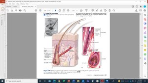
The arrector pili is a small band of smooth muscle attached to the hair follicle. It is responsible for piloerection (goose bumps) and is absent in eyebrows, eyelashes, and some facial hairs.
Nails
Structure and Function of Nails
Fingernails and toenails are hard, transparent epidermal derivatives produced by the nail matrix. They are composed of densely packed keratinized cells, providing strength and resistance to flaking. The nail bed and matrix are formed from invaginated epidermis and consist of the stratum basale and stratum spinosum. Melanocytes exist in the nail matrix but typically do not produce melanin, so nails remain clear.
Functions: Protection, support for gripping, tactile sensitivity, clinical indicators
Nail matrix: Responsible for nail growth
Nail body: Slides distally over the nail bed
Burns
Classification and Clinical Implications
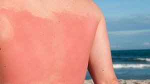
Burns are a leading cause of accidental death, with common causes including fires, hot water, sunlight, radiation, chemicals, and electrical shock. Deaths result mainly from fluid loss, infection, and toxic effects of eschar. The severity of burns is classified by degree:
First-Degree Burns: Affect only the epidermis; symptoms include redness, mild swelling, pain, and dryness. Healing occurs within a week without scarring.
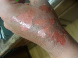
Second-Degree Burns: Affect both epidermis and dermis; symptoms include redness, swelling, pain, and blisters. Healing typically occurs within 2-3 weeks, with possible scarring.
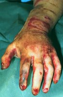
Third-Degree Burns: Affect all skin layers and may reach underlying tissues. Symptoms include white, black, or charred appearance and numbness. Healing requires medical intervention and skin grafts, with significant scarring.
Skin Cancer
Types and Clinical Evaluation
Skin cancer is common, with most cases caused by ultraviolet (UV) radiation damaging DNA in epidermal cells. Major types include:
Basal cell carcinoma: Most common and least dangerous
Squamous cell carcinoma
Merkel cell carcinoma: Rare but aggressive
Melanoma: Deadliest form, arising from melanocytes
Clinicians use the ABCDE rule to evaluate suspicious pigmented lesions.
Gland Type | Location | Secretion | Function |
|---|---|---|---|
Merocrine (Eccrine) | Palms, soles, forehead | Watery sweat | Thermoregulation |
Apocrine | Axillae, areolae, pubic region | Viscous sweat | Scent, body odor |
Sebaceous | Face, scalp, chest, back | Sebum | Lubrication, protection |
Ceruminous | External auditory canal | Cerumen | Protection, antimicrobial |
Mammary | Breast tissue | Milk | Nutrition |
Additional info: The notes expand on brief points with academic context, including definitions, examples, and clinical relevance. The table summarizes gland types for comparison.
