 Indietro
The Integumentary System: Structure, Function, and Clinical Relevance
Guida di studio - Note intelligenti
 Appunti personalizzati basati sui tuoi materiali, ampliati con definizioni chiave, esempi e contesto.
The Integumentary System
Overview and Components
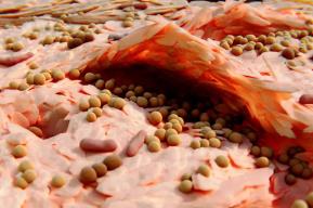
The integumentary system is a complex organ system comprised of the skin, hair, nails, subcutaneous tissue, and various glands. It serves as the body's primary interface with the external environment, providing protection, sensation, temperature regulation, and synthesis of essential vitamins.
Skin: The largest organ, acting as a barrier and regulator.
Hair: Provides insulation and protection against dust and foreign particles.
Nails: Enhance fingertip sensation and protect the distal phalanges.
Subcutaneous tissue: Connects skin to underlying structures and provides insulation.
Glands: Include sweat and sebaceous glands, crucial for thermoregulation and lubrication.
Functions of the Integumentary System
Protection: Prevents entry of harmful substances and fluid loss.
Insulation: Hair and subcutaneous fat help maintain body temperature.
Sensation: Skin contains nerve endings for touch, temperature, and pain.
Vitamin Synthesis: Skin synthesizes vitamin D upon exposure to sunlight.


Skin Structure and Layers
General Properties
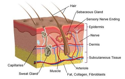
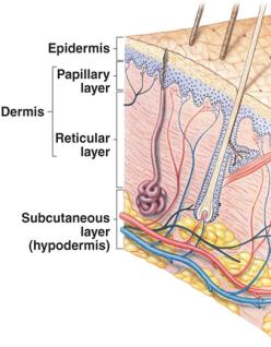
The skin is a large, resilient organ, typically 2–3 mm thick, with a high density of sweat glands, blood vessels, melanocytes, and nerve endings per square inch. 
Cutaneous Membrane
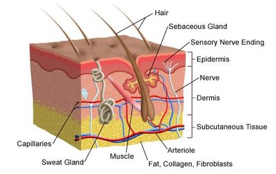
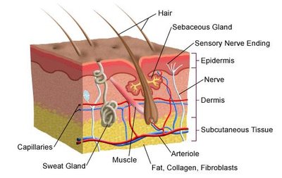
The skin consists of two main layers:
Epidermis: The outer, avascular layer made of stratified squamous keratinizing epithelium.
Dermis: The inner layer composed of connective tissue, housing nerves, blood vessels, glands, and hair follicles.
Hypodermis (Subcutaneous Tissue)
The hypodermis is a deep layer connecting skin to muscles and bones, providing padding and insulation.
Epidermis: Structure and Cell Types
The epidermis is dominated by keratinocytes, which produce keratin, a tough protein. It is nourished by diffusion from the dermis and contains melanocytes (pigment-producing cells) and Langerhans cells (immune function). 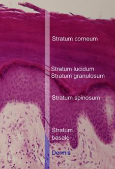
Thin vs. Thick Skin
Thin Skin: Covers most of the body, has four layers of keratinocytes.
Thick Skin: Found on palms and soles, has a fifth layer (stratum lucidum) and a thicker stratum corneum.


Strata of the Epidermis
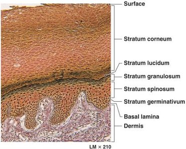
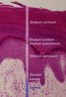
The epidermis in thick skin is organized into five strata, from deep to superficial:
Stratum germinativum (basale)
Stratum spinosum
Stratum granulosum
Stratum lucidum (only in thick skin)
Stratum corneum

Stratum Germinativum / Basale
Contains stem cells (basal cells) responsible for mitosis.
Forms epidermal ridges (fingerprints) with the papillary layer of the dermis.
Only living layer undergoing cell division.
Specialized Cells of the Epidermis
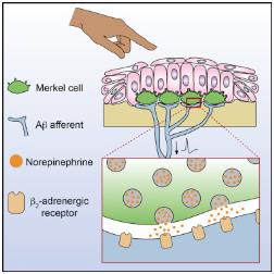
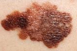
Merkel cells: Respond to touch, triggering the nervous system.
Melanocytes: Produce melanin, which absorbs UV radiation and protects DNA.

Epidermal Pigmentation
Melanin: Provides pigmentation and UV protection.
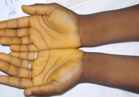
Carotene: Orange-yellow pigment, most evident in light-skinned individuals, can be converted to vitamin A.


Stratum Spinosum
Produced by division of basal cells.
Contains Langerhans cells, active in immune response.
Stratum Granulosum
Cells stop dividing and produce keratin and keratohyalin granules.
Granules promote dehydration and cross-linking of keratin filaments.
Stratum Lucidum
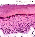
Clear layer found only in thick skin.
Stratum Corneum
Exposed surface, 15–30 layers of keratinized cells.
Water resistant, dry, and unsuitable for microorganism growth.
Cells are shed and replaced every two weeks.
Dermis
Structure and Function
The dermis lies beneath the epidermis and contains nerves, blood vessels, hair follicles, smooth muscle, glands, and lymphatic tissue. It is composed of loose connective tissue, including collagen, elastin, and reticular fibers. 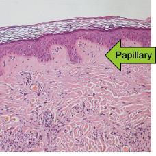
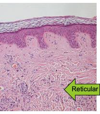
Layers of the Dermis
Papillary layer: Outermost, extends blood vessels toward the epidermis, forms papillary ridges (fingerprints).
Reticular layer: Dense, continuous with hypodermis, contains most structures (sweat glands, hair cells).
Main Roles of Skin
Protection
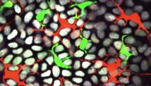
The skin acts as an anatomical barrier, with overlapping layers of dead cells and immune cells (Langerhans and dendritic T cells) defending against pathogens. 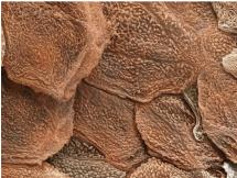
Sensation
The skin contains nerve endings and specialized cells (Merkel cells) that detect temperature, touch, pressure, vibration, and injury. 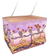
Temperature Regulation
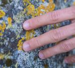
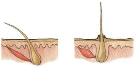
Skin regulates temperature through blood vessel dilation/constriction and arrector pili muscle activity. Blood flow is prioritized for vital organs during cold exposure. 


Vitamin D Production
Sunlight enables epidermal cells to convert cholesterol-related steroids into cholecalciferol (vitamin D3), which is processed by the liver and kidneys to form calcitriol, essential for calcium and phosphorus absorption. 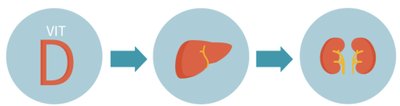
Vitamin D Deficiencies
Deficiency can lead to rickets, osteopenia, osteoporosis, cardiovascular disease, cognitive impairment, severe asthma, cancer, and mood disorders. 

Hypodermis
Structure and Function
The hypodermis is not classified as a skin layer but attaches skin to muscle and bone, supplies blood vessels and nerves, and contains fat for padding and insulation.
Hair
Types and Functions
Lanugo: Fine, unpigmented hair on fetuses.
Vellus: Short, unpigmented "peach fuzz" hair.
Terminal: Fully developed, longer, coarser, and pigmented hair.

Arrector Pili Muscles
Located in the dermis, these muscles contract to pull hair upright, aiding in heat conservation. 
Nails
Structure and Function
Nails are made of keratin and serve as protective plates and enhance fingertip sensation. 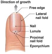
Sweat Glands
Eccrine (Merocrine) Glands
Produce watery sweat (99% water) for temperature regulation.
Distributed throughout the body, concentrated on palms, soles, forehead, and armpits.
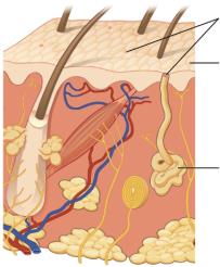
Apocrine Glands
Larger, located in armpits, groin, and breasts.
Produce concentrated secretions after puberty, associated with body odor.

Sebaceous Glands
Structure and Function
Sebaceous glands secrete sebum, an oily substance that lubricates and waterproofs skin and hair, inhibiting microorganism growth. 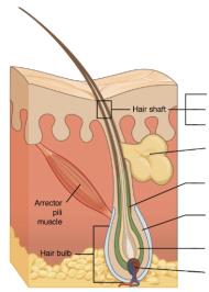
Acne Vulgaris
Acne occurs when sebum and dead skin cells plug hair follicles, leading to inflammation and infection. Treatments include benzoyl peroxide, antibacterial soap, anti-inflammatory drugs, antibiotics, vitamin A derivatives, and topical antiseptics. 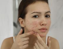
Inflammation and Regeneration
Inflammatory Response
Inflammation is a protective response to injury or infection, characterized by redness, pain, heat, swelling, and loss of function.
Vasodilation: Increases blood flow and permeability, allowing defensive materials to enter.
Phagocyte Migration: Macrophages ingest and destroy foreign matter.
Nutrient Release: Supports defensive cells and tissue repair.
Fibrin Formation: Forms a clot to localize infection.
Pus Formation: Mixture of dead cells and debris, cleared by the circulatory system or forms an abscess.
Regeneration
The final phase involves fibroblast activity, collagen fiber formation, and ground substance production. Repairs do not restore original condition; hair follicles, sweat glands, and muscle tissue are replaced by fibrous tissue (fibrosis).
Scar Tissue: Inflexible, non-cellular tissue forms after injury.
Keloids: Overgrowth of collagen tissue, influenced by genetics.
Layer | Main Features |
|---|---|
Epidermis | Stratified squamous keratinizing epithelium, avascular, contains melanocytes and Langerhans cells |
Dermis | Loose connective tissue, blood vessels, nerves, glands, hair follicles |
Hypodermis | Subcutaneous fat, attaches skin to muscle/bone, insulation |
Stratum | Function |
|---|---|
Germinativum (Basale) | Stem cell division, attachment to dermis |
Spinosum | Immune response, cell division |
Granulosum | Keratin and keratohyalin production |
Lucidum | Clear layer, only in thick skin |
Corneum | Keratinized, water-resistant, protective |
Gland Type | Location | Function |
|---|---|---|
Eccrine | Entire body | Watery sweat, temperature regulation |
Apocrine | Armpits, groin, breasts | Concentrated sweat, body odor |
Sebaceous | Throughout skin except palms/soles | Oily sebum, lubrication, waterproofing |
Key Equations
Vitamin D Synthesis:
Additional info:
Recent research suggests vitamin D may help prevent and treat diabetes, hypertension, glucose intolerance, multiple sclerosis, and depression.
Scarless healing in fetuses is an area of ongoing research.
