 Indietro
The Muscle System: Structure, Function, and Major Muscles
Guida di studio - Note intelligenti
 Appunti personalizzati basati sui tuoi materiali, ampliati con definizioni chiave, esempi e contesto.
The Muscle System
Relationship Between Body Movement and Muscle Action
The muscular system is essential for producing movement in the human body. Muscles contract to generate force, which is transmitted to bones via tendons, resulting in movement at joints. The efficiency and type of movement depend on muscle action, fiber type, and the arrangement of muscle groups.
Aerobic (endurance) exercise increases muscle capillaries, mitochondria, and myoglobin synthesis, leading to greater endurance and resistance to fatigue.
Resistance (anaerobic) exercise causes muscle hypertrophy, increasing fiber size, mitochondria, myofilaments, glycogen stores, and connective tissue, resulting in greater strength and size.
Disuse atrophy occurs when muscles are inactive, leading to degeneration and loss of mass.
Force of Muscle Contractions
Depends on the number of cross bridges formed, affected by:
Number of muscle fibers stimulated (recruitment)
Relative size of fibers (hypertrophy increases force)
Frequency of stimulation
Degree of muscle stretch (optimal sarcomere length: 80–120% resting length)
Muscle Fiber Types
Classified by speed of contraction (slow or fast) and metabolic pathway (oxidative or glycolytic).
Load affects contraction speed and duration: muscles contract fastest with no load; greater load slows contraction and shortens duration.
Characteristics of Smooth Muscle Tissue
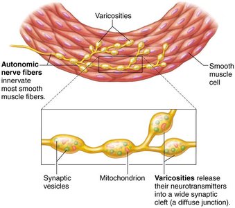
Smooth muscle is found in the walls of most hollow organs (except the heart). It is responsible for involuntary movements such as peristalsis and regulation of blood vessel diameter.
Contains two layers:
Longitudinal layer: fibers run parallel to the organ's long axis; contraction shortens the organ.
Circular layer: fibers run around the organ; contraction constricts the lumen.
Alternating contractions mix and propel substances through the organ.
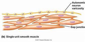
Innervated by the autonomic nervous system; contains varicosities (bulbous nerve swellings) that release neurotransmitters into diffuse junctions.
No striations or sarcomeres; contains overlapping thick and thin filaments.
Cells are electrically coupled by gap junctions, allowing synchronized contractions.
Regulation of contraction is controlled by nerves, hormones, or local chemical changes.
Muscle Attachments: Origin and Insertion
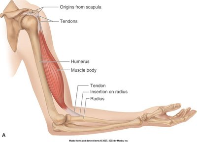
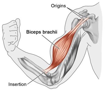
Muscles attach to bones at two points: the origin (stationary during contraction) and the insertion (moves during contraction). Movement occurs as the insertion is pulled toward the origin.
Origin is usually proximal; insertion is distal.
Muscles can only pull, not push.
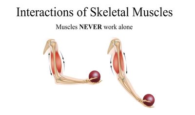
Muscle groups work in opposition: what one group does, another undoes.
Functional Groups of Muscles
Prime mover (agonist): major responsibility for a specific movement.
Antagonist: opposes or reverses a movement.
Synergist: assists prime movers, adds force, reduces unwanted movement.
Fixator: stabilizes the origin of the prime mover.
Naming Muscles
Muscles are named based on location, shape, size, direction of fibers, number of origins, location of attachments, and action.
Examples:
Temporalis (location: temporal bone)
Deltoid (shape: triangle)
Biceps (number of origins: two)
Sternocleidomastoid (attachments: sternum, clavicle, mastoid process)
Flexor or extensor (action)
Fascicle Arrangements
The arrangement of fascicles determines muscle shape and function.
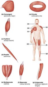
Circular: concentric rings (e.g., orbicularis oris)
Convergent: broad origin, fascicles converge to a single tendon (e.g., pectoralis major)
Parallel: fascicles parallel to long axis (e.g., sartorius)
Pennate: short fascicles attach obliquely to a central tendon (e.g., rectus femoris)
Levers in Muscle Movement
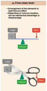
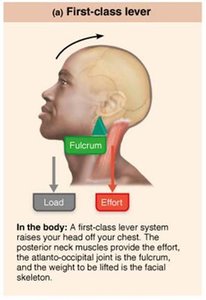
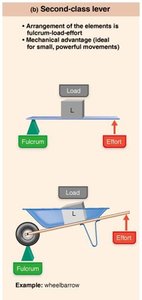
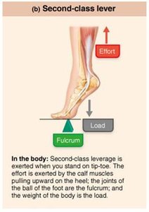
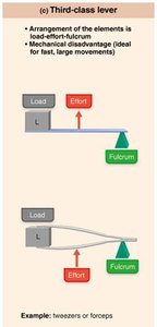
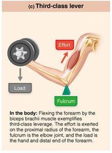
Most skeletal muscles move bones using leverage. The lever system consists of a rigid bar (bone), a fulcrum (joint), effort (muscle contraction), and load (resistance).
Effort farther from fulcrum than load = mechanical advantage.
Effort nearer to fulcrum than load = mechanical disadvantage.
Lever Class | Arrangement | Example |
|---|---|---|
First-class | Load-Fulcrum-Effort | Neck muscles raising head |
Second-class | Fulcrum-Load-Effort | Standing on tiptoe |
Third-class | Load-Effort-Fulcrum | Biceps flexing forearm |
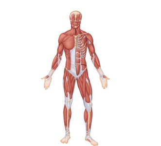
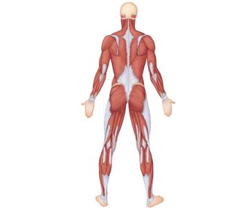
Major Muscles: Points of Attachment and Function
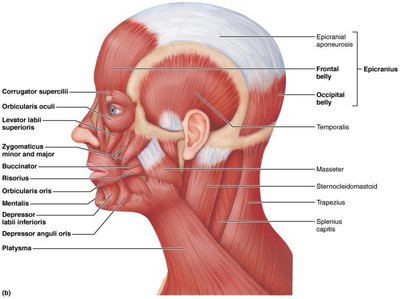
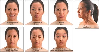
Muscles of Facial Expression
Facial muscles insert into skin, allowing for nonverbal communication. They are divided into muscles of the scalp and face.
Muscles of Mastication and Tongue Movement
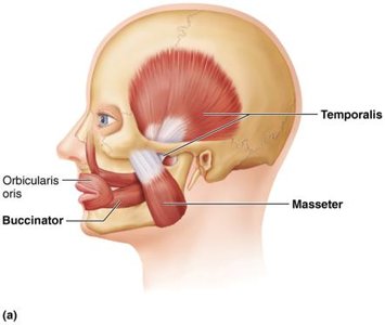
Jaw closure: temporalis, masseter
Grinding: pterygoids
Chewing: buccinator
Tongue movement: genioglossus, hyoglossus, styloglossus
Neck Muscles
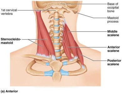
Sternocleidomastoid divides neck into anterior and posterior triangles.
Muscles of swallowing: tongue, buccinator, pharyngeal muscles.
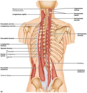
Neck movement: anterolateral neck muscles; trunk extension and posture: erector spinae, quadratus lumborum.
Muscles of Breathing
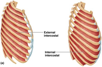
Inspiratory: diaphragm, external intercostals (enlarge rib cage)
Expiratory: internal intercostals, abdominal muscles (decrease rib cage size)
Abdominal Wall Muscles
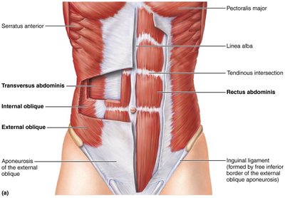
Rectus abdominis, external obliques, internal obliques, transversus abdominis
Actions: lateral flexion, trunk rotation, assist in urination, defecation, childbirth, vomiting, coughing, screaming
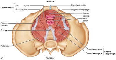
Pelvic Diaphragm Muscles
Functions: seals inferior outlet, supports organs, lifts pelvic floor, resists intra-abdominal pressure
Shoulder Muscles
Extrinsic shoulder muscles fix and move the scapula for increased arm movement range.
Actions: elevation, depression, rotation, lateral/medial movement, protraction, retraction
Groups: anterior thorax, posterior thorax
Arm Muscles
Prime movers: pectoralis major, latissimus dorsi, deltoid
Rotator cuff: supraspinatus, infraspinatus, teres minor, subscapularis, coracobrachialis, teres major
Forearm Muscles
Anterior: biceps brachii, brachialis, brachioradialis (flex and supinate forearm)
Posterior: triceps brachii (forearm extension)
Thigh Muscles
Anterior: flex femur at hip, extend leg at knee (iliopsoas, tensor fasciae latae, rectus femoris, sartorius)
Posterior: extend thigh, flex leg (hamstrings: biceps femoris, semitendinosus, semimembranosus)
Medial: adduct thigh (adductor magnus, adductor longus, adductor brevis, pectineus, gracilis)
Quadriceps Femoris
Four heads: rectus femoris, vastus lateralis, vastus medialis, vastus intermedius
Insert into quadriceps tendon, patella, tibial tuberosity
Powerful knee extensor
Gluteal Muscles
Abductors and rotators: gluteus maximus, medius, minimus, piriformis, obturator externus/internus, gemellus
Leg Muscles
Anterior compartment: tibialis anterior, extensor digitorum longus, extensor hallucis longus (toe extension, ankle dorsiflexion)
Posterior compartment: soleus, tibialis posterior, gastrocnemius (ankle plantar flexion)
Summary Table: Major Muscle Groups and Functions
Region | Muscles | Function |
|---|---|---|
Head | Facial, mastication, tongue | Expression, chewing, swallowing |
Neck | Sternocleidomastoid, scalene, erector spinae | Head movement, posture |
Thorax | Intercostals, diaphragm | Breathing |
Abdomen | Rectus abdominis, obliques, transversus abdominis | Trunk movement, abdominal pressure |
Pelvis | Levator ani, coccygeus | Pelvic support |
Shoulder | Pectoralis major, deltoid, rotator cuff | Arm movement |
Arm | Biceps, triceps, brachialis | Forearm movement |
Thigh | Quadriceps, hamstrings, adductors | Hip/knee movement |
Leg | Tibialis anterior, gastrocnemius, soleus | Ankle/toe movement |
Key Equations
Muscle force generation:
Mechanical advantage:
Additional info:
Muscle groups are often named using multiple criteria for clarity and specificity.
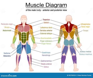
Tables and diagrams are used to clarify muscle locations, functions, and lever mechanics.
