 Indietro
The Reproductive System: Functional Anatomy and Physiology
Guida di studio - Note intelligenti
 Appunti personalizzati basati sui tuoi materiali, ampliati con definizioni chiave, esempi e contesto.
The Reproductive System
Overview
The reproductive system is responsible for producing, storing, nourishing, and transporting gametes. It consists of gonads, ducts, accessory glands/organs, and external genitalia. Gonads produce gametes and hormones: testes produce sperm and androgens, while ovaries produce oocytes and estrogen/progesterone.
Cell Division in Reproduction
Mitosis
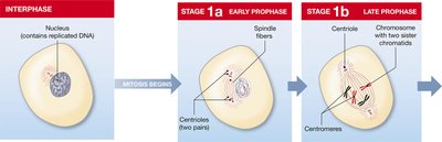
Mitosis is the process by which somatic cells divide, resulting in two genetically identical diploid cells (46 chromosomes). It is essential for growth, repair, and maintenance of tissues.
Prophase: Chromosomes condense, spindle fibers form.
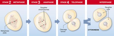
Metaphase: Chromosomes align at the metaphase plate.
Anaphase: Chromatids separate to opposite poles.
Telophase: Chromosomes uncoil, nucleus reforms.
Cytokinesis: Division of cytoplasm, forming two daughter cells.
Meiosis
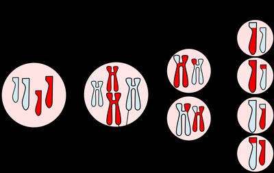
Meiosis is the process by which gametes are produced, involving two rounds of cell division (meiosis I and II). It results in four haploid cells (23 chromosomes each), ensuring genetic diversity.
Meiosis I: Homologous chromosomes pair (synapsis) and exchange genetic material (crossing-over).
Meiosis II: Sister chromatids separate, forming haploid gametes.
Key Differences: Synapsis and crossing-over occur only in meiosis, not mitosis.
Male Reproductive System
Functional Anatomy
The male reproductive system produces sperm and androgens. It includes the testes, duct system, accessory glands, and penis.
Testes: Male gonads, held in the scrotum, lined by serous membrane.
Dartos muscle: Wrinkles scrotal skin.
Cremaster muscle: Pulls testes closer to the body for temperature regulation (90°F - 95°F).
Tunica albuginea: Fibrous covering, forms septa dividing testis into lobules.
Seminiferous tubules: Site of sperm formation.
Leydig cells: Produce testosterone.
Nurse (Sertoli) cells: Support sperm formation.
Spermatogenesis
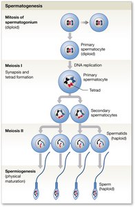
Spermatogenesis is the process of sperm formation, involving mitosis, meiosis, and spermiogenesis (maturation). It begins at puberty and occurs in the seminiferous tubules.
Spermatogonia: Undergo mitosis to form primary spermatocytes.
Primary spermatocytes: Undergo meiosis to form spermatids (haploid).
Nurse cells: Provide nutrients, chemical stimuli, and produce inhibin.
Sperm Structure
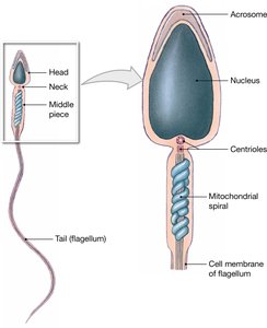
A sperm cell has four regions: head, neck, middle piece, and tail. The head contains the acrosome, which has enzymes to penetrate the ovum. Sperm must absorb nutrients from surrounding fluid.
Head: Contains nucleus and acrosome.
Neck: Contains centrioles.
Middle piece: Contains mitochondria for energy.
Tail (flagellum): Provides motility.
Male Reproductive Tract and Accessory Glands
Sperm mature as they pass through the reproductive tract: seminiferous tubules → epididymis → vas deferens → ejaculatory duct → urethra → penis. Accessory glands (seminal, prostate, bulbo-urethral) contribute fluids to semen, activating sperm and providing nutrients.
Seminal glands: 60% of semen volume, secrete fructose, prostaglandins, fibrinogens.
Prostate: 20-30% of semen volume, secretes acidic fluid with enzymes.
Bulbourethral glands: Produce thick alkaline mucus for lubrication.
External Male Genitalia
The penis conducts urine and semen to the external environment. It contains three columns of erectile tissue: two corpora cavernosa and one corpus spongiosum (surrounds urethra).
Glans penis: Covered by prepuce.
Scrotum: Houses testes.
Male Reproductive Hormones
The hypothalamus releases GnRH, stimulating the anterior pituitary to release FSH and LH. FSH targets nurse cells for spermatogenesis; LH targets Leydig cells for testosterone production.
FSH: Promotes spermatogenesis, produces inhibin.
LH: Promotes testosterone production, maintains libido, bone, and muscle growth.
Female Reproductive System
Functional Anatomy
The female reproductive system produces sex hormones and gametes (ova), protects and supports embryos, and nourishes newborns. It includes ovaries, uterus, vagina, external genitalia, and breasts.
Ovaries: Female gonads, produce oocytes, estrogen, progesterone, and inhibin.
Ligaments: Ovarian and suspensory ligaments connect ovaries to uterus and pelvic wall.
Oogenesis
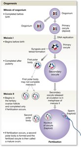
Oogenesis is the process of oocyte formation, beginning before birth and continuing monthly between puberty and menopause. Oogonia complete mitosis before birth, forming primary oocytes that begin meiosis and pause at prophase I until puberty.
Meiosis I: Produces secondary oocytes and polar bodies.
Meiosis II: Completed only after fertilization.
Ovarian follicles: Primordial, primary, secondary, and tertiary follicles support oocyte development.
Corpus luteum: Releases progesterone after ovulation.
Corpus albicans: Degenerating corpus luteum (scar tissue).
Ovarian Cycle
The ovarian cycle describes cyclical events in the ovaries: follicular phase (FSH stimulates follicle development), ovulation (LH surge releases oocyte), and luteal phase (corpus luteum releases progesterone).
Follicular phase: FSH stimulates tertiary follicle development.
Ovulation: LH surge triggers release of secondary oocyte.
Luteal phase: Corpus luteum releases progesterone; degenerates if not fertilized.
Uterine Tubes and Uterus
The uterine tubes are the site of fertilization. Fimbriae help catch oocytes, which are transported by ciliary movements and peristalsis. The uterus protects and nourishes the embryo, with a thick muscular layer for contractions during labor.
Endometrium: Inner layer, functional layer undergoes cyclical changes.
Myometrium: Muscular middle layer.
Cervix: Opening to uterus.
Uterine Cycle (Menstrual Cycle)
The uterine cycle ranges from 21-35 days and includes three phases:
Menstrual phase: Shedding of endometrial functional layer, triggered by decline in progesterone.
Proliferative phase: Endometrium rebuilds, stimulated by rising estrogen.
Secretory phase: Uterine glands secrete nutrients, stimulated by estrogen and progesterone.
Female Reproductive Hormones
GnRH from the hypothalamus stimulates FSH and LH release. LH promotes progesterone secretion by corpus luteum; FSH promotes follicle development. Estrogen and progesterone regulate the cycle and maintain reproductive organs.
Estrogen: Stimulates bone/muscle growth, maintains sex characteristics.
Progesterone: Prepares uterus for pregnancy.
Inhibin: Inhibits FSH secretion.
Female Reproductive Cycle
The female reproductive cycle coordinates ovarian and uterine cycles through hormones. Negative feedback inhibits LH secretion when estrogen is low; positive feedback promotes FSH, LH, and GnRH when estrogen rises. LH surge triggers ovulation; corpus luteum produces progesterone, maintaining endometrium.
GnRH: Causes LH & FSH secretion.
Estrogen: Rebuilds endometrium.
LH surge: Triggers ovulation.
Progesterone: Maintains endometrium; decline causes menstruation.
HCG: Maintains corpus luteum if fertilization occurs.
Vagina and External Female Genitalia
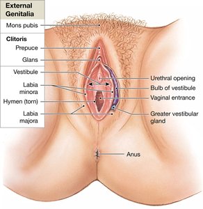
The vagina is an elastic tube connecting the uterus to the exterior, serving as a passageway for menstrual fluids, sperm, and birth canal. The vulva includes labia minora, labia majora, clitoris, and vestibular glands for lubrication.
Labia minora: Protect vagina and urethra.
Clitoris: Erectile tissue, comparable to corpora cavernosa.
Greater vestibular glands: Lubricate vaginal entrance.
Mammary Glands
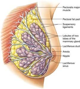
Mammary glands produce milk to nourish newborns. Breast milk contains nutrients and antibodies. Lactation is controlled by hormones: prolactin (production), oxytocin (ejection), and chorionic somatomammotropin (placenta). Colostrum is the initial milk, rich in nutrients and IgA antibodies.
Lobules: Contain secretory alveoli.
Lactiferous ducts: Merge into lactiferous sinus near nipple.
Areola: Pigmented area around nipple.
Key Formulas and Concepts
Chromosome Number: Diploid (somatic cells): 46 chromosomes; Haploid (gametes): 23 chromosomes.
Negative Feedback: Progesterone inhibits GnRH, LH, FSH secretion.
Positive Feedback: Estrogen promotes GnRH, LH, FSH secretion.
Example: During the menstrual cycle, a surge in LH triggers ovulation, while a decline in progesterone causes menstruation.
Additional info: The notes include expanded explanations of cell division, reproductive anatomy, and hormonal regulation to ensure completeness and academic quality for exam preparation.
