 Indietro
The Skeletal System: Bone Tissue – Structure, Function, and Physiology
Guida di studio - Note intelligenti
 Appunti personalizzati basati sui tuoi materiali, ampliati con definizioni chiave, esempi e contesto.
The Skeletal System: Bone Tissue
Overview and Functions of the Skeletal System
The skeletal system is a complex organ system composed of bones, cartilage, dense connective tissue, adipose, and nervous tissue. It provides the structural framework for the body and supports various physiological processes essential for life.
Support: Bones provide the rigid framework for the body and anchor soft tissues and organs.
Protection: The skeleton protects vital organs such as the brain (skull), spinal cord (vertebrae), and thoracic organs (rib cage).
Movement: Bones act as levers for muscles, enabling movement.
Mineral Homeostasis: Bones store minerals, primarily calcium and phosphorus, and release them into the bloodstream as needed.
Blood Cell Formation: Hematopoiesis occurs in red marrow cavities, producing red and white blood cells and platelets.
Triglyceride Storage: Yellow marrow stores fat as an energy reserve.
Hormone Production: Osteocalcin, produced by bones, regulates insulin secretion, glucose levels, and metabolism.
Types of Bones in the Skeleton
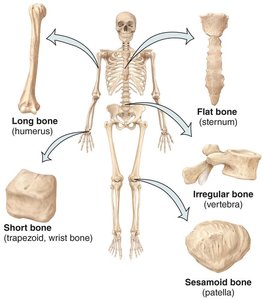
Bones are classified by shape and structure, each adapted for specific functions in the body.
Long bones: Longer than they are wide (e.g., humerus, femur).
Short bones: Nearly equal in length and width (e.g., carpals, tarsals).
Flat bones: Thin and often curved (e.g., sternum, ribs, skull bones).
Irregular bones: Complex shapes (e.g., vertebrae, some facial bones).
Sesamoid bones: Develop within tendons (e.g., patella).
Structure of a Long Bone
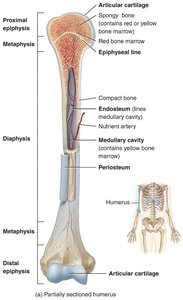
Long bones have a characteristic structure that supports their function in movement and weight-bearing.
Diaphysis: The shaft or central part of the bone.
Epiphyses: The ends of the bone, which articulate with other bones.
Metaphyses: The regions between the diaphysis and epiphyses; contain the epiphyseal (growth) plate in growing bones.
Articular cartilage: Hyaline cartilage covering the epiphyses at joints, reducing friction and absorbing shock.
Periosteum: Dense connective tissue membrane covering the diaphysis, containing osteogenic cells for growth and repair.
Medullary cavity: Hollow space within the diaphysis, containing yellow marrow in adults.
Endosteum: Thin membrane lining the medullary cavity, containing bone-forming cells.
Bone Tissue Composition
Bones are composed of a specialized connective tissue with a robust extracellular matrix and several cell types.
Extracellular matrix: Approximately 15% water, 30% collagen fibers (providing flexibility), and 55% crystallized mineral salts (mainly hydroxyapatite, providing hardness).
Hydroxyapatite: Crystals formed from calcium phosphate and calcium hydroxide, giving bone its strength.
Bone Cells
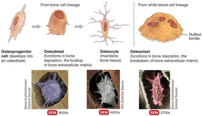
Bone tissue contains four main types of cells, each with distinct roles in bone formation, maintenance, and remodeling.
Osteoprogenitor cells: Stem cells that differentiate into osteoblasts.
Osteoblasts: Bone-building cells that secrete bone matrix and initiate calcification.
Osteocytes: Mature bone cells that maintain bone tissue.
Osteoclasts: Large cells that resorb (break down) bone, releasing calcium into the blood.
Compact vs. Spongy Bone
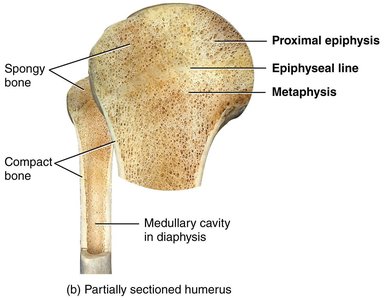
Bones contain two types of osseous tissue: compact bone and spongy bone, each with unique structural and functional properties.
Compact bone: Dense and strong, forming the outer layer of bones; provides protection and support.
Spongy bone (cancellous or trabecular bone): Lightweight, with a porous structure; found mainly in the epiphyses and inside flat bones; supports and protects red bone marrow.
Blood and Nerve Supply of Bone
Bones are highly vascularized and innervated, ensuring their vitality and ability to remodel and repair.
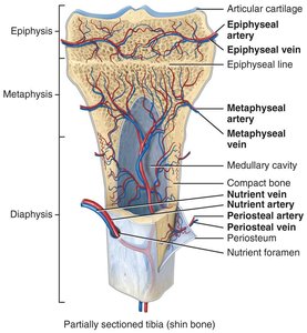
Periosteal arteries and veins: Supply the outer compact bone via Volkmann’s canals.
Nutrient artery and vein: Enter the diaphysis through the nutrient foramen, supplying the inner compact bone and medullary cavity.
Metaphyseal and epiphyseal arteries and veins: Supply the metaphyses and epiphyses, respectively.
Bone Formation and Growth
Ossification (Osteogenesis)
Ossification is the process of bone formation, which occurs during embryonic development, bone growth, remodeling, and fracture repair.
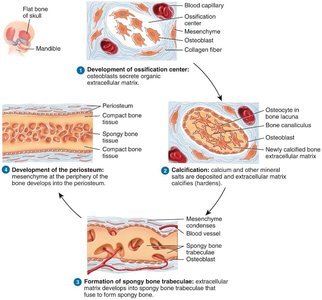
Intramembranous ossification: Bone develops directly from mesenchymal tissue; forms flat bones like the skull and clavicle.
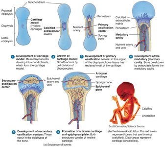
Endochondral ossification: Bone forms by replacing hyaline cartilage; forms most bones of the body, especially long bones.
Steps of Endochondral Ossification
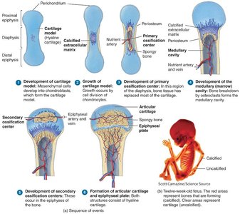
Endochondral ossification involves a series of steps that transform cartilage into bone.
Bone collar forms around the diaphysis of the cartilage model.
Central cartilage in the diaphysis calcifies, then develops cavities.
Periosteal bud invades cavities, forming spongy bone.
Diaphysis elongates, and the medullary cavity forms; secondary ossification centers appear in the epiphyses.
Epiphyses ossify; hyaline cartilage remains only in the epiphyseal plates and articular cartilages.
Growth in Bone Length: Epiphyseal Plate
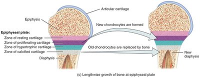
Long bones grow in length at the epiphyseal plate through interstitial growth, involving several distinct zones.
Resting (quiescent) zone: Inactive cartilage on the epiphyseal side.
Proliferation (growth) zone: Rapidly dividing chondrocytes push the epiphysis away from the diaphysis.
Hypertrophic zone: Older chondrocytes enlarge and erode, forming spaces.
Calcification zone: Matrix calcifies, chondrocytes die.
Ossification (osteogenic) zone: New bone forms as osteoblasts cover calcified spicules.
Growth in Bone Thickness (Appositional Growth)
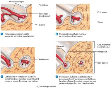
Bones increase in diameter through appositional growth, involving the coordinated actions of osteoblasts and osteoclasts.
Osteoblasts deposit new bone on the outer surface.
Osteoclasts resorb bone from the inner surface, widening the medullary cavity.
Bone Remodeling and Repair
Bone Remodeling
Bone remodeling is a continuous process where old bone is replaced by new bone tissue, maintaining bone strength and mineral homeostasis.
Involves bone resorption by osteoclasts and bone deposition by osteoblasts.
Remodeling is influenced by mechanical stress, hormones, and nutritional status.
Bone Fractures and Repair
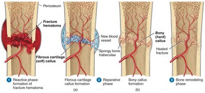
Bone fractures are breaks in bone continuity, classified by their characteristics. Bone repair occurs in three main phases:
Reactive phase: Formation of a fracture hematoma (inflammatory response).
Reparative phase: Formation of a fibrocartilaginous (soft) callus, followed by a bony (hard) callus.
Bone remodeling phase: Remodeling of the bony callus to restore normal bone structure.
Common Types of Bone Fractures
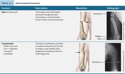
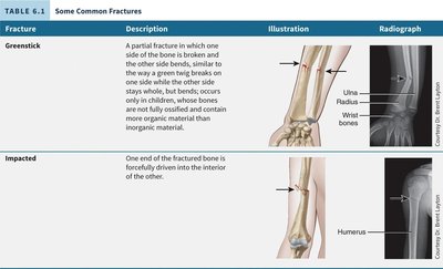
Fractures are classified based on the nature of the break:
Fracture | Description |
|---|---|
Open (Compound) | The broken ends of the bone protrude through the skin. |
Comminuted | The bone is splintered, crushed, or broken into pieces. |
Greenstick | A partial fracture in which one side of the bone is broken and the other side bends; common in children. |
Impacted | One end of the fractured bone is forcefully driven into the interior of the other. |
Calcium Homeostasis and Bone
Bones play a critical role in maintaining calcium balance in the body. Calcium is essential for muscle contraction, nerve function, and blood clotting.
Parathyroid hormone (PTH): Released when blood calcium levels are low; stimulates osteoclasts to resorb bone and release calcium.
Calcitriol (active vitamin D): Increases calcium absorption from the intestines.
Calcitonin: Released by the thyroid gland; inhibits osteoclast activity, promoting calcium deposition in bone.
Factors Affecting Bone Growth and Remodeling
Minerals
Calcium and phosphorus: Essential for bone matrix hardness.
Magnesium, fluoride, manganese: Important for matrix formation and enzyme activation.
Vitamins
Vitamin A: Stimulates osteoblast activity.
Vitamin C: Required for collagen synthesis.
Vitamin D: Promotes calcium absorption; deficiency leads to rickets or osteomalacia.
Vitamins K and B12: Needed for protein synthesis in bone.
Hormones
Growth hormone (GH): Stimulates bone growth via IGFs.
Thyroid hormones (T3, T4): Promote bone growth.
Insulin: Increases bone protein synthesis.
Sex hormones (estrogen, testosterone): Stimulate osteoblasts, promote growth spurt, and regulate bone remodeling.
Parathyroid hormone (PTH): Increases bone resorption.
Calcitonin: Inhibits bone resorption.
Exercise, Aging, and Bone Health
Effect of Exercise
Weight-bearing activities stimulate osteoblasts, increasing bone mass and strength. Regular exercise helps prevent bone loss with aging.
Aging and Bone Tissue
Bone mass increases during growth, stabilizes in adulthood, and decreases with age, especially in postmenopausal women due to reduced estrogen levels. This increases the risk of osteoporosis.
Bone Disorders and Homeostatic Imbalances
Osteoporosis: Bone resorption exceeds formation, leading to decreased bone mass and increased fracture risk; most common in postmenopausal women.
Rickets: Inadequate calcification of bone matrix in children, causing bone deformities.
Osteomalacia: Softening of bones in adults due to poor mineralization.
Osteoarthritis: Degeneration of articular cartilage, causing joint pain and stiffness.
Osteomyelitis: Infection of bone, often by bacteria.
Osteopenia: Reduced bone mass below normal but not low enough to be classified as osteoporosis.
Osteosarcoma: Malignant bone tumor, primarily affecting osteoblasts.
Key Medical Terminology
Osteoblasts: Cells that build new bone.
Osteoclasts: Cells that break down bone tissue.
Periosteum: Connective tissue membrane covering the diaphysis of long bones.
Epiphyseal plate: Growth plate in long bones, responsible for lengthwise growth.
Sample Questions for Review
Name the two types of bone found in the skeletal system.
__________ help create new bone, and __________ help destroy old bone. Both are key for bone remodeling processes. Choices: Osteoclasts/Osteoblasts/Osteocytes
This type of fracture results in the bone protruding through the skin.
What is the name of the connective tissue that surrounds the diaphysis?
How many bones are in the adult human skeleton?
