 Indietro
Urinary System: Structure, Function, and Physiology
Guida di studio - Note intelligenti
 Appunti personalizzati basati sui tuoi materiali, ampliati con definizioni chiave, esempi e contesto.
The Urinary System
Organs and Functions
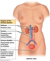
The urinary system is essential for the removal of metabolic wastes, regulation of blood volume and pressure, and maintenance of homeostasis. It consists of paired kidneys, ureters, the urinary bladder, and the urethra.
Kidneys: Produce urine and regulate blood composition.
Ureters: Transport urine from kidneys to bladder.
Urinary bladder: Temporarily stores urine prior to elimination.
Urethra: Conducts urine to the exterior; also transports semen in males.
Urination (Micturition)
Urination is the process of eliminating urine from the body. It involves contraction of the muscular bladder, forcing urine through the urethra.
Excretion: Removal of metabolic wastes from body fluids.
Elimination: Discharge of wastes from the body.
Homeostatic regulation: Regulates blood volume, blood pressure, plasma ion concentration, pH, conserves nutrients, and assists in detoxification.
The Kidneys
Location and Structure
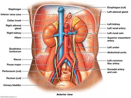
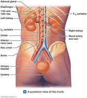
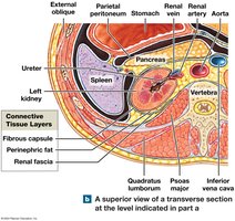
The kidneys are retroperitoneal organs, with the left kidney slightly superior to the right. Each kidney is held in position by the peritoneum, adjacent organs, and connective tissues.
Fibrous capsule: Collagen layer covering the kidney.
Perinephric fat: Adipose tissue cushioning the kidney.
Renal fascia: Dense outer layer anchoring the kidney.
Internal Anatomy
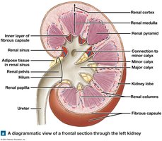
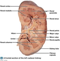
Hilum: Medial indentation for entry/exit of vessels and ureter.
Renal sinus: Internal cavity containing adipose tissue and urine collecting ducts.
Renal cortex: Superficial region.
Renal medulla: Inner region with renal pyramids.
Renal columns: Tissue bands between pyramids.
Minor calyx: Collects urine from renal papilla.
Major calyx: Formed by merging minor calyces.
Renal pelvis: Funnel-shaped chamber draining urine into ureter.
Blood Supply
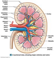
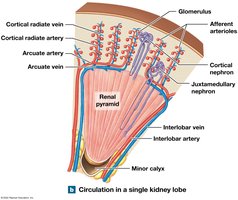
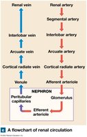
Kidneys receive 20–25% of cardiac output. Blood enters via the renal artery, branches into segmental, interlobar, arcuate, and cortical radiate arteries, and is delivered to nephrons via afferent arterioles. Venous return follows cortical radiate, arcuate, and interlobar veins, draining into the renal vein.
Nephron Structure
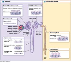
The nephron is the functional unit of the kidney, responsible for urine production. It consists of:
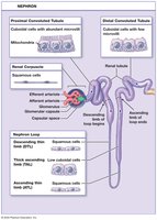
Renal corpuscle: Filters blood; includes glomerular (Bowman's) capsule and glomerulus.
Renal tubule: Proximal convoluted tubule (PCT), nephron loop (Loop of Henle), distal convoluted tubule (DCT).
Renal Corpuscle
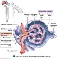
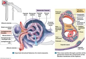
Glomerular capsule: Outer wall; contains glomerular capillaries.
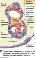
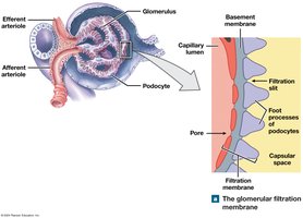
Podocytes: Specialized cells with foot processes forming filtration slits.
Glomerulus: Ball of capillaries; blood enters via afferent arteriole, leaves via efferent arteriole.
Filtration membrane: Fenestrated endothelium, basement membrane, filtration slits.
Renal Tubule Segments
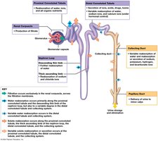
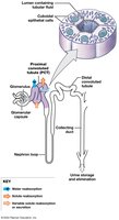
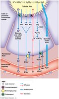
PCT: Cuboidal epithelium with microvilli; reabsorbs nutrients, ions, water.
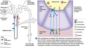
Nephron loop: Descending limb (permeable to water), ascending limb (permeable to solutes).
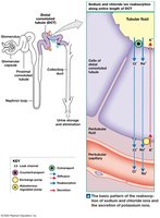
DCT: Cuboidal cells lacking microvilli; selective reabsorption and secretion.
Juxtaglomerular Complex (JGC)
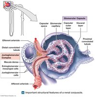
Macula densa: Chemoreceptors/baroreceptors in DCT.
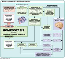
Juxtaglomerular cells: Baroreceptors; secrete renin.
Extraglomerular mesangial cells: Feedback control between macula densa and juxtaglomerular cells.
Collecting System
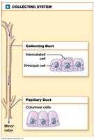
Collecting ducts: Receive fluid from nephrons; principal cells reabsorb water, intercalated cells regulate acid–base balance.
Papillary duct: Drains into minor calyx.
Nephron Types
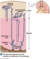
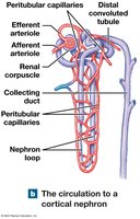
Cortical nephrons: 85% of nephrons; short loops, peritubular capillaries.
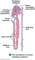
Juxtamedullary nephrons: Long loops, vasa recta; important for urine concentration.
Renal Physiology
Urine Formation
Urine formation involves three main processes:
Filtration: Blood pressure forces water and solutes across glomerular capillaries into the capsular space.
Reabsorption: Movement of water and solutes from filtrate back to blood.
Secretion: Transport of solutes from blood into filtrate.
Glomerular Filtration
Driven by hydrostatic pressure.
Filtration membrane: Restricts cells and proteins, allows water and small solutes.
Filtration Pressures
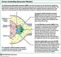
Glomerular hydrostatic pressure (GHP): Pushes water and solutes out of blood.
Capsular hydrostatic pressure (CsHP): Opposes GHP.
Net hydrostatic pressure (NHP):
Blood colloid osmotic pressure (BCOP): Pulls water back into capillaries.
Net filtration pressure (NFP):
Glomerular Filtration Rate (GFR)
GFR: Amount of filtrate produced per minute; regulated by autoregulation, hormonal, and autonomic mechanisms.
Hormonal Regulation
Renin–angiotensin–aldosterone system (RAAS): Increases blood pressure and GFR via vasoconstriction and fluid retention.
Natriuretic peptides (ANP): Increase GFR and urine production, decrease blood volume.
Reabsorption and Secretion
Transport Mechanisms
Diffusion, osmosis, leak channels, carrier-mediated transport: Includes facilitated diffusion, active transport, cotransport, and countertransport.
Transport maximum: Saturation point for carrier proteins; excess substrate appears in urine.
Renal threshold: Plasma concentration at which a compound appears in urine (e.g., glucose, amino acids).
Segment Functions
Proximal Convoluted Tubule (PCT)
Reabsorbs: Organic nutrients, ions, water.
Secretes: Nitrogenous wastes, drugs.
Sodium reabsorption: Via diffusion, cotransport, countertransport, and sodium-potassium pumps.
Nephron Loop
Descending limb: Permeable to water, not solutes.
Ascending limb: Impermeable to water, reabsorbs sodium and chloride.
Distal Convoluted Tubule (DCT)
Selective reabsorption: Sodium, calcium.
Active secretion: Ions, acids, drugs, toxins.
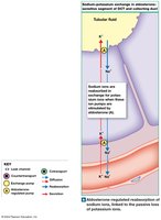
Hormonal effects: Aldosterone increases sodium reabsorption, parathyroid hormone regulates calcium.
Collecting System
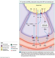
Reabsorption: Sodium (aldosterone), water (ADH), bicarbonate.
Secretion: Hydrogen ions (low pH), bicarbonate (high pH).
Regulating Urine Volume and Concentration
Countercurrent Multiplication
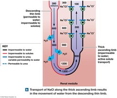
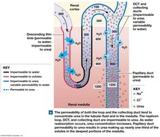
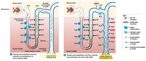
Countercurrent multiplication in the nephron loop creates a medullary osmotic gradient, allowing concentration of urine.
Descending limb: Permeable to water.
Ascending limb: Active transport of Na+, K+, Cl-; impermeable to water.
Water Reabsorption
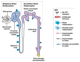
Obligatory: Occurs in PCT and descending limb; cannot be prevented.
Facultative: Occurs in DCT and collecting duct; regulated by ADH.
Vasa Recta and Countercurrent Exchange
Vasa recta: Returns solutes and water to circulation, maintains medullary gradient.
Summary of Juxtamedullary Nephron Function
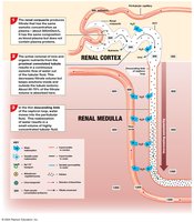
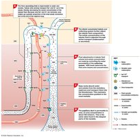
Step 1: Filtration at renal corpuscle.
Step 2: Reabsorption at PCT.
Step 3: Water reabsorption at descending limb.
Step 4: Solute reabsorption at ascending limb.
Step 5: Selective reabsorption/secretion at DCT and collecting system.
Step 6: Final adjustments in collecting system.
Step 7: Vasa recta maintains gradient.
Step 8: Urea contributes to medullary gradient.
Normal Urine Composition
Clear, sterile liquid.
Yellow color: Due to urobilin.
Urinalysis: Diagnostic tool for kidney function.
Kidney Function Tests
Creatinine clearance: Estimates GFR.
BUN (blood urea nitrogen): Measures urea in blood.
Urine Transport, Storage, and Elimination
Ureters
Muscular tubes: Transport urine from kidneys to bladder.
Layers: Mucosa (transitional epithelium), muscular layer (smooth muscle), connective tissue.
Peristaltic contractions: Move urine every 30 seconds.
Urinary Bladder
Hollow, muscular organ: Temporary urine storage.
Stabilized: By peritoneal folds and ligaments.
Rugae: Folds that disappear as bladder fills.
Trigone: Funnels urine into urethra.
Neck: Contains internal urethral sphincter (involuntary control).
Detrusor muscle: Contracts to expel urine.
Urethra
Female: Short (3–5 cm), extends from bladder to vestibule.
Male: Long (18–20 cm), passes through prostate, perineal muscle, and penis.
External urethral sphincter: Skeletal muscle, voluntary control.
Urinary Reflexes
Urine storage reflex: Involves spinal reflexes and pontine storage center; inhibits detrusor contraction, contracts sphincters.
Urine voiding reflex: Initiated when bladder fills; detrusor contracts, sphincters relax.
Effects of Aging
Nephrolithiasis: Formation of kidney stones.
Decrease in functional nephrons, GFR, sensitivity to ADH.
Problems with urinary reflexes: Loss of muscle tone, incontinence, urinary retention (especially in males).
Integration with Other Systems
The urinary system interacts with the integumentary, respiratory, and digestive systems to maintain body fluid composition and excrete wastes.
