 Indietro
A Tour of the Cell: Structure, Function, and Diversity
Guida di studio - Note intelligenti
 Appunti personalizzati basati sui tuoi materiali, ampliati con definizioni chiave, esempi e contesto.
Introduction to the Cell
The Cell as the Fundamental Unit of Life
Cells are the basic structural and functional units of all living organisms. Life emerges at the cellular level, and all organisms are composed of one or more cells. The similarities among all cells demonstrate the unity of life, while differences reveal its diversity.
All organisms are made of cells
Cells are the fundamental unit of life
Unity and diversity: Similarities show common ancestry; differences reflect adaptation and specialization
Types of Cells
Prokaryotic vs. Eukaryotic Cells
Cells are classified into two main types: prokaryotic and eukaryotic. This distinction is based on structural and functional differences.
Prokaryotic Cells: Found in Bacteria and Archaea; lack membrane-bound organelles and a nucleus.
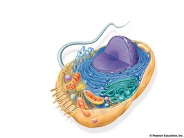
Eukaryotic Cells: Found in Eukarya (protists, fungi, animals, plants); possess membrane-bound organelles and a nucleus.
Basic features of all cells:
Plasma membrane
Semifluid cytosol
Chromosomes (genetic material)
Ribosomes (protein synthesis)
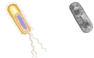
Prokaryotic Cell Structure
No membrane-bound organelles
No nucleus; DNA is in the nucleoid region
Cytoplasm is bound by the plasma membrane
Eukaryotic Cell Structure
DNA is enclosed within a nucleus
Contains membrane-bound organelles
Cytoplasm is the region between the plasma membrane and nucleus
Generally larger than prokaryotic cells
Cell Size and Surface Area
Limits to Cell Size
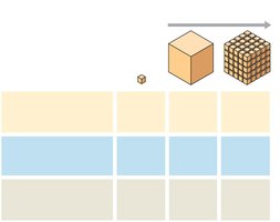
Metabolic requirements set upper limits on cell size. The plasma membrane is critical for the exchange of materials. As a cell increases in size, its volume grows faster than its surface area, limiting efficient exchange.
Surface area-to-volume ratio is a key factor in cell size and function.
Internal Membranes and Compartmentalization
Membrane Structure and Function
In eukaryotic cells, internal membranes partition the cell into organelles, allowing specialized environments for different functions. Biological membranes are composed of a double layer of phospholipids with embedded proteins.
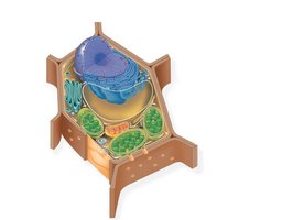
Plant and animal cells share most organelles
Animal, Plant, and Fungal Cells
Comparative Cell Types
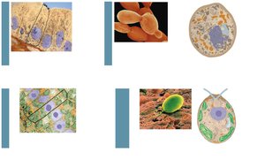
Animal, plant, and fungal cells are all eukaryotic but have distinct features. For example, plant cells have cell walls and chloroplasts, while animal cells do not.
The Nucleus
Structure and Function
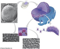
The nucleus contains most of the cell's genetic material (DNA) and is surrounded by a double-membrane nuclear envelope. It is easily identifiable and typically about 5 μm in diameter.
Nuclear envelope: Double membrane with pore complexes for molecular transport
Nuclear lamina: Protein network supporting nuclear shape
Nuclear matrix: Internal framework (hypothetical)
Chromosomes: DNA-protein complexes (chromatin) that condense during cell division
Nucleolus: Site of ribosomal RNA (rRNA) synthesis and ribosome assembly
Ribosomes
Structure and Function
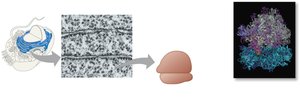
Ribosomes are complexes of rRNA and protein that carry out protein synthesis. They are not membrane-bound and are found in both prokaryotes and eukaryotes.
Free ribosomes: Suspended in cytosol; synthesize cytosolic proteins
Bound ribosomes: Attached to ER or nuclear envelope; synthesize membrane-bound or secretory proteins
The Endomembrane System
Components and Functions
The endomembrane system regulates protein traffic and performs metabolic functions. It includes the nuclear envelope, endoplasmic reticulum (ER), Golgi apparatus, lysosomes, vesicles, vacuoles, and plasma membrane.
Components are connected directly or via vesicles
Highly diverse in structure and function
Endoplasmic Reticulum (ER)
Smooth ER: Lacks ribosomes; synthesizes lipids, metabolizes carbohydrates, detoxifies drugs, stores calcium
Rough ER: Studded with ribosomes; produces secretory proteins and membranes
Golgi Apparatus
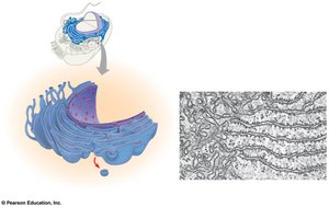
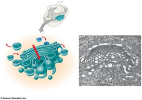
The Golgi apparatus modifies, sorts, and packages products from the ER. It consists of flattened membranous sacs called cisternae.
Cis face: Receiving side, near ER
Trans face: Shipping side, away from ER
Products are modified as they move through the Golgi
Vesicles transport materials to and from the Golgi
Lysosomes
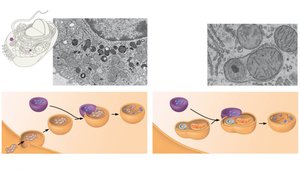
Lysosomes are membranous sacs of hydrolytic enzymes that digest macromolecules in animal cells. They function best in acidic environments and are involved in intracellular digestion (phagocytosis) and recycling (autophagy).
Vacuoles
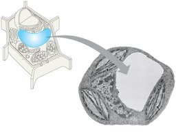
Vacuoles are large vesicles with diverse functions, including storage, waste disposal, and maintaining cell turgor in plants. Central vacuoles in plant cells store water and organic compounds.
Mitochondria and Chloroplasts
Energy Conversion Organelles
Mitochondria and chloroplasts are involved in energy conversion. Mitochondria are the sites of cellular respiration, generating ATP using oxygen. Chloroplasts, found in plants and algae, are the sites of photosynthesis, converting solar energy to chemical energy.
Endosymbiont Theory
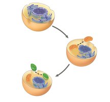
This theory proposes that mitochondria and chloroplasts originated as free-living prokaryotes engulfed by ancestral eukaryotic cells. Evidence includes their double membranes, own DNA, and ribosomes.
Peroxisomes
Oxidative Organelles
Peroxisomes are organelles that carry out oxidation reactions, breaking down fatty acids and detoxifying harmful substances. They produce hydrogen peroxide (H2O2) as a by-product, which is then converted to water.
The Cytoskeleton
Structure and Function
The cytoskeleton is a network of fibers that organizes cell structure and activities. It provides support, anchors organelles, and enables cell movement.
Microtubules: Thick, hollow rods; involved in cell shape, chromosome movement, and organelle transport
Microfilaments: Thin, solid rods of actin; involved in cell shape, muscle contraction, and movement
Intermediate filaments: Intermediate size; provide structural support and anchor organelles
Extracellular Components
Cell Walls in Plants
Plant cells have cell walls made of cellulose, providing protection, structural support, and preventing excessive water uptake. Cell walls may have multiple layers, and plasmodesmata connect adjacent cells.
Extracellular Matrix (ECM) in Animals
The ECM is a network of glycoproteins (e.g., collagen, proteoglycans, fibronectin) that supports animal cells, facilitates communication, and influences cell behavior through integrin receptors.
Cell Junctions
Types of Cell Junctions
Cells interact and communicate through specialized junctions:
Plasmodesmata (plants): Channels allowing cytoplasmic exchange between cells
Tight junctions (animals): Prevent leakage of extracellular fluid
Desmosomes (animals): Anchor cells together
Gap junctions (animals): Allow passage of ions and small molecules between cells
