 Indietro
Chapter 6
Guida di studio - Note intelligenti
 Appunti personalizzati basati sui tuoi materiali, ampliati con definizioni chiave, esempi e contesto.
6.1 Biologists Use Microscopes and Biochemistry to Study Cells
History of Cell Discovery
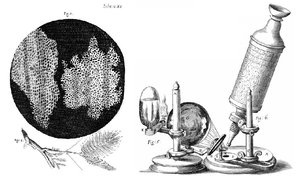
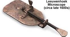
Early cell studies were made possible by the invention of microscopes. Robert Hooke first described 'cells' in cork, while Anton van Leeuwenhoek observed living cells in pond water using a simple microscope.
Robert Hooke: Observed dead cork cells, coined the term 'cell'.
Anton van Leeuwenhoek: Improved microscope design, observed living microorganisms.
Types of Microscopes
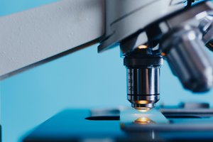
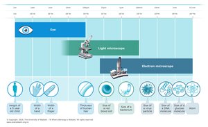
Light Microscope (LM): Uses visible light and glass lenses to magnify specimens up to 1,000x. Suitable for viewing living cells, but cannot resolve structures smaller than 0.2 micrometers.
Electron Microscope (EM): Uses electron beams and magnetic lenses for much higher resolution (up to 2 nm). Cannot be used on living specimens.
Transmission Electron Microscope (TEM): Electrons pass through thin specimens, revealing internal structures.
Scanning Electron Microscope (SEM): Electrons scan the surface, producing detailed 3D images of cell surfaces.
Microscope Resolution and Specimen Size
Resolution is the ability to distinguish two points as separate. Electron microscopes provide much higher resolution than light microscopes, allowing visualization of much smaller structures.
Cell Fractionation
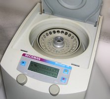
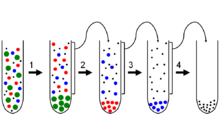
Cell fractionation uses a centrifuge to separate cellular components by mass, allowing study of individual organelles.
Centrifuge: Spins samples at high speed to separate components into pellets and supernatant.
Application: Enables biochemical analysis of specific cell parts.
6.2 Eukaryotic Cells Have Internal Membranes That Compartmentalize Their Functions
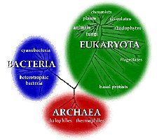
Prokaryotes vs. Eukaryotes: Overview
Cells are classified as prokaryotic or eukaryotic based on structural differences.
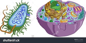
Prokaryotes: Bacteria and Archaea; lack membrane-bound organelles; DNA in nucleoid; generally smaller.
Eukaryotes: Animals, plants, fungi, protists; have membrane-bound organelles; DNA in nucleus; generally larger.
Common Features of All Cells
Plasma membrane
Genetic material (chromosomes)
Cytosol (liquid part of cytoplasm)
Ribosomes (protein synthesis)
Key Differences Between Prokaryotes and Eukaryotes
Feature | Prokaryotes | Eukaryotes |
|---|---|---|
DNA Location | Nucleoid (no membrane) | Nucleus (membrane-bound) |
Organelles | Absent | Present (membrane-bound) |
DNA Structure | Circular | Linear |
Size | Smaller | Larger |
Surface Area to Volume Ratio
Cells must maintain a high surface area to volume ratio to efficiently exchange materials with their environment. As cells grow, volume increases faster than surface area, limiting size.
Diffusion: Movement of molecules from high to low concentration; efficient in small cells with large surface area relative to volume.
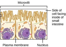
6.3 The Eukaryotic Cell’s Genetic Instructions Are Housed in the Nucleus and Carried Out by the Ribosomes
Nucleus
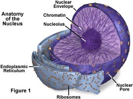
The nucleus is the control center of the cell, containing most of the cell's DNA. It is surrounded by a double membrane called the nuclear envelope, which contains nuclear pores for molecular transport.
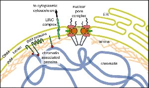
Nuclear envelope: Double membrane with pores.
Nuclear lamina: Protein network supporting the envelope.
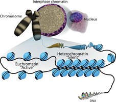
Chromatin: DNA wrapped around histone proteins; condenses into chromosomes during cell division.
Nucleolus: Site of ribosomal RNA (rRNA) synthesis and ribosome assembly.
Ribosomes
Ribosomes are complexes of rRNA and proteins that synthesize proteins. They are not membrane-bound and can be found free in the cytosol or bound to the endoplasmic reticulum (ER).
Free ribosomes: Synthesize proteins for use within the cell.
Bound ribosomes: Attached to ER; synthesize proteins for membranes, lysosomes, or export.
6.4 The Endomembrane System Regulates Protein Traffic and Performs Metabolic Functions
Components of the Endomembrane System
Nuclear envelope
Endoplasmic reticulum (ER): Smooth and rough
Golgi apparatus
Vesicles and vacuoles
Plasma membrane
Vesicles
Vesicles are small, membrane-bound sacs that transport materials within, into, or out of the cell.
Endoplasmic Reticulum (ER)
Smooth ER: Synthesizes lipids, carbohydrates, steroids, and hormones; detoxifies chemicals; regulates calcium storage.
Rough ER: Studded with ribosomes; synthesizes proteins, which are folded and transported in vesicles to the Golgi apparatus.
Golgi Apparatus
The Golgi apparatus modifies, sorts, and packages proteins and lipids for storage or transport out of the cell. It consists of flattened sacs called cisternae.
Cis face: Receiving side
Trans face: Shipping side
Lysosomes
Lysosomes are membrane-bound vesicles containing digestive enzymes. They break down macromolecules, recycle cell components (autophagy), and digest engulfed particles (phagocytosis).
Vacuoles
Food vacuoles: Formed by phagocytosis in animal cells.
Central vacuole: Found in plant cells; stores water, salts, and other substances.
6.5 Mitochondria and Chloroplasts Change Energy from One Form to Another
Mitochondria
Mitochondria are the sites of cellular respiration, converting glucose and oxygen into ATP, the cell's energy currency.
Double membrane structure
Inner membrane forms cristae to increase surface area
Matrix contains enzymes for respiration
Contain their own DNA and ribosomes
Chloroplasts
Chloroplasts are found in plants and algae and are the sites of photosynthesis, converting light energy, water, and CO2 into glucose and oxygen.
Double membrane
Thylakoids (stacked into grana) contain chlorophyll
Stroma is the fluid surrounding thylakoids
Contain their own DNA and ribosomes
Endosymbiotic Theory
This theory proposes that mitochondria and chloroplasts originated as free-living prokaryotes engulfed by ancestral eukaryotic cells. Evidence includes their double membranes, circular DNA, and independent reproduction.
Peroxisomes
Peroxisomes are specialized vesicles that detoxify harmful substances by transferring hydrogen to oxygen, forming hydrogen peroxide, which is then converted to water and oxygen.
6.6 The Cytoskeleton Is a Network of Fibers That Organizes Structures and Activities in the Cell
Functions of the Cytoskeleton
Provides mechanical support and maintains cell shape
Anchors organelles
Facilitates cell movement and intracellular transport
Types of Cytoskeletal Fibers
Type | Structure | Function |
|---|---|---|
Microtubules | Thick, hollow rods (tubulin) | Support, tracks for organelle movement, cell division (centrioles), cilia & flagella |
Microfilaments | Thin, solid rods (actin) | Cell shape, muscle contraction, cell movement (pseudopodia) |
Intermediate Filaments | Medium-sized, various proteins | Structural support, more permanent, nuclear lamina |
6.7 Extracellular Components and Connections Between Cells Help Coordinate Cellular Activities
Plant Cell Wall
The cell wall provides protection and structural support. It is composed mainly of cellulose microfibrils. The primary wall is thin and flexible, while the secondary wall is thicker and more rigid. The middle lamella, rich in pectin, glues adjacent cells together.
Extracellular Matrix (ECM) of Animal Cells
The ECM is composed of glycoproteins (such as collagen), proteoglycans, and fibronectin. It provides structural support, helps cells adhere, and transmits signals via integrins (transmembrane proteins) connected to the cytoskeleton.
Cell Junctions
Plasmodesmata (plants): Channels connecting plant cells, allowing cytosol and molecules to pass between cells.
Tight junctions (animals): Seal cells together, preventing leakage of extracellular fluid.
Desmosomes (animals): Anchor cells together at spots, providing mechanical stability.
Gap junctions (animals): Channels allowing ions and small molecules to pass between cells, similar to plasmodesmata in plants.
