 Indietro
Cell Communication and Cell Cycle Study Guide (General Biology)
Guida di studio - Note intelligenti
 Appunti personalizzati basati sui tuoi materiali, ampliati con definizioni chiave, esempi e contesto.
Q1. Match the type of cell signaling:
Background
Topic: Cell Communication
This question tests your understanding of different types of cell signaling, including how cells communicate over short and long distances.
Key Terms:
Endocrine signaling: Long-distance signaling via hormones in the bloodstream.
Synaptic signaling: Short-distance signaling between neurons using neurotransmitters.
Step-by-Step Guidance
Read each description and identify whether it refers to signaling across long distances or between neighboring cells.
Recall that hormones typically travel through the bloodstream for long-distance communication (endocrine signaling).
Remember that neurons communicate with each other via synaptic signaling, which involves neurotransmitters crossing a synapse.
Try solving on your own before revealing the answer!
Final Answer:
Signaling across long distances: Endocrine signaling
Electrical signal triggering neurotransmitter release: Synaptic signaling
Endocrine signaling uses hormones in the bloodstream, while synaptic signaling is specific to neurons.
Q2. What are the three steps of cell signaling? What happens during each step?
Background
Topic: Cell Signaling Pathways
This question tests your knowledge of the basic steps in cell signaling: reception, transduction, and response.
Key Terms:
Reception: The cell detects a signaling molecule from the outside.
Transduction: The signal is relayed and amplified inside the cell.
Response: The cell reacts to the signal, often by changing its activity.
Step-by-Step Guidance
Identify the first step, where the cell receives the signal (reception).
Describe the second step, where the signal is passed through a series of molecules (transduction).
Explain the third step, where the cell makes a change in response to the signal (response).
Try solving on your own before revealing the answer!
Final Answer:
Reception: Cell detects signaling molecule.
Transduction: Signal is relayed and amplified.
Response: Cell changes activity in response.
These steps ensure that cells can respond appropriately to external signals.
Q3. What are the three major types of membrane receptors?
Background
Topic: Membrane Receptors
This question tests your knowledge of the main classes of membrane receptors involved in cell signaling.
Key Terms:
G-protein coupled receptors (GPCRs)
Receptor tyrosine kinases (RTKs)
Ion channel receptors
Step-by-Step Guidance
Recall the three main types of membrane receptors.
Think about their functions: GPCRs activate G proteins, RTKs phosphorylate tyrosines, and ion channels allow ions to pass.
Write the names in the blanks provided.
Try solving on your own before revealing the answer!
Final Answer:
1. G-protein coupled receptor (GPCR)
2. Receptor tyrosine kinase (RTK)
3. Ion channel receptor
These are the three major types of membrane receptors in cell signaling.
Q4. Properly order the steps of signaling via a G-protein coupled receptor
Background
Topic: GPCR Signaling Pathway
This question tests your understanding of the sequence of events in G-protein coupled receptor signaling.
Key Terms:
Ligand: A molecule that binds to a receptor.
GDP/GTP: Guanine nucleotides involved in G protein activation.
Enzyme: Triggers cellular response.
Step-by-Step Guidance
Identify the step where the ligand binds to the GPCR, activating the G protein.
Next, the activated G protein dissociates from the receptor and activates an enzyme.
The enzyme triggers a cascade that leads to a cellular response.
Finally, the G protein hydrolyzes GTP to GDP, becoming inactive.
Try solving on your own before revealing the answer!
Final Answer:
Ligand binds to GPCR outside of the cell, causing the G protein to switch GDP for GTP and become activated.
Activated G protein dissociates from the GPCR and activates an enzyme.
Enzyme triggers a cascade that causes a cellular response.
G protein hydrolyzes GTP back to GDP, releasing the enzyme and becoming inactivated.
This is the correct order for GPCR signaling.
Q5. What is a second messenger? What is its purpose?
Background
Topic: Second Messengers in Cell Signaling
This question tests your understanding of the role of second messengers in amplifying and relaying signals inside the cell.
Key Terms:
Second messenger: Small molecules that transmit signals from receptors to target molecules inside the cell.
Amplification: Increasing the strength of the signal.
Step-by-Step Guidance
Define what a second messenger is.
Explain its purpose: to relay and amplify the signal inside the cell.
Think of examples like cAMP, Ca2+, and IP3.
Try solving on your own before revealing the answer!
Final Answer:
A second messenger is a small molecule that relays signals from receptors on the cell surface to target molecules inside the cell, amplifying the signal and triggering a response.
Q6. Give three examples of second messengers.
Background
Topic: Second Messengers
This question tests your ability to recall common second messengers used in cell signaling pathways.
Key Terms:
cAMP (cyclic AMP)
Ca2+ (calcium ions)
IP3 (inositol triphosphate)
Step-by-Step Guidance
Recall the most common second messengers.
Write down three examples.
Try solving on your own before revealing the answer!
Final Answer:
cAMP
Ca2+
IP3
These are widely used second messengers in cell signaling.
Q7. A group of enzymes called __________ add a phosphate group to a protein, while another group of enzymes called __________ remove a phosphate group from a protein.
Background
Topic: Protein Modification in Signaling
This question tests your knowledge of enzymes involved in phosphorylation and dephosphorylation.
Key Terms:
Kinases: Add phosphate groups.
Phosphatases: Remove phosphate groups.
Step-by-Step Guidance
Recall the names of enzymes that add and remove phosphate groups.
Fill in the blanks with the correct terms.
Try solving on your own before revealing the answer!
Final Answer:
Kinases add phosphate groups.
Phosphatases remove phosphate groups.
These enzymes regulate protein activity in signaling pathways.
Q8. True or false: adding a phosphate group ALWAYS results in activation of a protein, while removing a phosphate group always results in deactivation of the protein.
Background
Topic: Regulation of Protein Activity
This question tests your understanding of how phosphorylation affects protein activity.
Key Terms:
Phosphorylation: Addition of a phosphate group.
Dephosphorylation: Removal of a phosphate group.
Step-by-Step Guidance
Consider whether phosphorylation always activates proteins.
Think about whether dephosphorylation always deactivates proteins.
Recall that the effect depends on the specific protein and context.
Try solving on your own before revealing the answer!
Final Answer:
False. Phosphorylation can either activate or deactivate a protein, depending on the protein and context.
Q9. The second messenger cAMP is made by the enzyme __________ hydrolyzing pyrophosphate from the molecule __________.
Background
Topic: cAMP Production
This question tests your knowledge of how cAMP is synthesized in cells.
Key Terms:
Adenylyl cyclase: Enzyme that makes cAMP.
ATP: Molecule used to make cAMP.
Step-by-Step Guidance
Recall the enzyme responsible for cAMP production.
Identify the molecule that is converted into cAMP.
Try solving on your own before revealing the answer!
Final Answer:
cAMP is made by adenylyl cyclase hydrolyzing pyrophosphate from ATP.
Q10. What area of the cell has a low Ca2+ concentration?
Background
Topic: Calcium Signaling
This question tests your knowledge of calcium ion distribution in cells.
Key Terms:
Cytosol: Fluid inside the cell.
Ca2+ concentration: Important for signaling.
Step-by-Step Guidance
Recall where Ca2+ is kept at low levels in the cell.
Think about the cytosol versus organelles like the ER.
Try solving on your own before revealing the answer!
Final Answer:
The cytosol has a low Ca2+ concentration.
Q11. The enzyme __________ cleaves a specific membrane lipid to generate DAG and PIP2 (which is converted to IP3).
Background
Topic: Lipid Signaling Pathways
This question tests your knowledge of enzymes involved in generating second messengers from membrane lipids.
Key Terms:
Phospholipase C: Enzyme that cleaves membrane lipids.
DAG and IP3: Second messengers.
Step-by-Step Guidance
Recall the enzyme responsible for cleaving PIP2.
Identify the products: DAG and IP3.
Try solving on your own before revealing the answer!
Final Answer:
Phospholipase C cleaves PIP2 to generate DAG and IP3.
Q12. What are the four key aspects of cell signaling?
Background
Topic: Cell Signaling Regulation
This question tests your knowledge of the main features that regulate cell signaling pathways.
Key Terms:
Amplification
Specificity
Scaffolding
Termination
Step-by-Step Guidance
Recall the four aspects: amplification, specificity, scaffolding, and termination.
Match each aspect to its description.
Try solving on your own before revealing the answer!
Final Answer:
Amplification: increases the strength of the signal and response
Specificity: ensures the response is appropriate for the signal
Scaffolding: enhancing a signal by scaffolding proteins that organize pathway components
Termination: stops the signal to reset the pathway
Q13. Controlled cell death is called __________.
Background
Topic: Apoptosis
This question tests your knowledge of the term for programmed cell death.
Key Terms:
Apoptosis: Controlled cell death.
Step-by-Step Guidance
Recall the term for controlled cell death.
Write the term in the blank.
Try solving on your own before revealing the answer!
Final Answer:
Apoptosis is controlled cell death.
Q14. Briefly explain the molecular mechanism of apoptosis in C. elegans.
Background
Topic: Apoptosis Mechanisms
This question tests your understanding of how apoptosis is regulated in the model organism C. elegans.
Key Terms:
Caspases: Proteins involved in apoptosis.
CED-3, CED-4, CED-9: Key genes in C. elegans apoptosis.
Step-by-Step Guidance
Recall the main proteins involved in apoptosis in C. elegans.
Describe how CED-9 inhibits apoptosis, while CED-4 and CED-3 promote it.
Explain the cascade that leads to cell death.
Try solving on your own before revealing the answer!
Final Answer:
In C. elegans, apoptosis is regulated by CED-9 (inhibitor), CED-4 (activator), and CED-3 (executioner caspase). When CED-9 is inactivated, CED-4 activates CED-3, leading to cell death.
Q15. What is a gamete? How many sets of chromosomes does a gamete have? How many sets of chromosomes does a somatic cell have?
Background
Topic: Chromosome Number in Cells
This question tests your understanding of the difference between gametes and somatic cells in terms of chromosome sets.
Key Terms:
Gamete: Sex cell (egg or sperm).
Somatic cell: Any body cell except gametes.
Haploid: One set of chromosomes.
Diploid: Two sets of chromosomes.
Step-by-Step Guidance
Define what a gamete is.
Recall that gametes are haploid (one set of chromosomes).
Somatic cells are diploid (two sets of chromosomes).
Try solving on your own before revealing the answer!
Final Answer:
Gamete: Sex cell (egg or sperm)
Gamete has one set of chromosomes (haploid)
Somatic cell has two sets of chromosomes (diploid)
Q16. The __________ is the narrow waist of a duplicated chromosome; the region where two sister chromatids are most tightly attached.
Background
Topic: Chromosome Structure
This question tests your knowledge of chromosome anatomy.
Key Terms:
Centromere: Region where chromatids are attached.
Step-by-Step Guidance
Recall the term for the region where sister chromatids are joined.
Write the term in the blank.
Try solving on your own before revealing the answer!
Final Answer:
Centromere is the narrow waist of a duplicated chromosome.
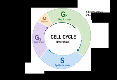
Q17. Label how many chromosomes and chromatids the cell would have at each phase of the cell cycle.
Background
Topic: Cell Cycle and Chromosome Number
This question tests your understanding of how chromosome and chromatid numbers change during the cell cycle.
Key Terms:
Chromosome: DNA molecule with genetic material.
Chromatid: One copy of a duplicated chromosome.
Cell cycle phases: G1, S, G2, M.
Step-by-Step Guidance
Review the cell cycle diagram and identify the phases: G1, S, G2, M.
Recall that in G1, the cell has unreplicated chromosomes (one chromatid per chromosome).
During S phase, DNA is replicated, so each chromosome now consists of two sister chromatids.
In G2 and M, the cell has duplicated chromosomes (two chromatids per chromosome).
After mitosis, each daughter cell returns to having single chromatids per chromosome.
Try solving on your own before revealing the answer!
Final Answer:
G1: Each chromosome has one chromatid.
S: DNA is replicated; chromosomes now have two chromatids.
G2: Chromosomes still have two chromatids.
M: Chromatids are separated; each daughter cell gets one chromatid per chromosome.
The cell cycle diagram helps visualize these changes.
Q18. Correctly name and order the steps of mitosis:
Background
Topic: Mitosis Stages
This question tests your knowledge of the sequence and description of mitosis stages.
Key Terms:
Prophase, Metaphase, Anaphase, Telophase, Cytokinesis
Step-by-Step Guidance
Recall the order of mitosis stages: prophase, metaphase, anaphase, telophase, cytokinesis.
Match each stage to its description (e.g., chromosomes align, chromatids separate).
Write the names in the correct order.
Try solving on your own before revealing the answer!
Final Answer:
Prophase: Nuclear envelope breaks down, chromosomes condense.
Metaphase: Chromosomes align at the cell equator.
Anaphase: Sister chromatids separate and migrate to opposite poles.
Telophase: Chromosomes de-condense, nuclear envelope reforms.
Cytokinesis: Cleavage furrow forms, cells separate.
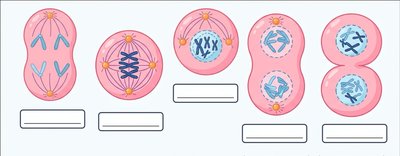
Q19. Label the stages of mitosis in the figure below:
Background
Topic: Mitosis Stages (Visual)
This question tests your ability to visually identify and label the stages of mitosis.
Key Terms:
Prophase, Metaphase, Anaphase, Telophase, Cytokinesis
Step-by-Step Guidance
Examine the diagram and identify the features of each stage (e.g., chromosome alignment, chromatid separation).
Recall the order and characteristics of mitosis stages.
Label each stage based on the visual cues.
Try solving on your own before revealing the answer!
Final Answer:
Prophase
Metaphase
Anaphase
Telophase
Cytokinesis
The diagram shows the sequence of mitosis stages.
