 Indietro
Cell Membrane Structure, Transport, and Eukaryotic Cell Organization: Study Notes for General Biology
Guida di studio - Note intelligenti
 Appunti personalizzati basati sui tuoi materiali, ampliati con definizioni chiave, esempi e contesto.
Cell Membrane Structure and the Fluid Mosaic Model
Overview of the Cell Membrane
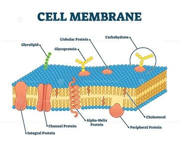
The cell membrane is a dynamic, flexible barrier that separates the interior of the cell from its external environment. It is composed of a phospholipid bilayer with embedded proteins, cholesterol, and carbohydrates, forming the fluid mosaic model.
Phospholipids: Amphipathic molecules with hydrophilic heads and hydrophobic tails, forming the bilayer.
Integral Proteins: Span the membrane, involved in transport and signaling.
Peripheral Proteins: Attached to the membrane surface, involved in cell signaling and structure.
Glycolipids and Glycoproteins: Lipids and proteins with carbohydrate chains, important for cell recognition.
Cholesterol: Stabilizes membrane fluidity across temperatures.
Fluid Mosaic Model
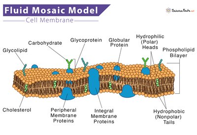
The fluid mosaic model describes the cell membrane as a fluid structure with a mosaic of proteins embedded in or associated with a phospholipid bilayer. This arrangement allows for flexibility, movement, and selective permeability.
Amphipathic Nature: Phospholipids have both hydrophilic and hydrophobic regions, enabling bilayer formation.
Membrane Proteins: Integral and peripheral proteins serve various functions, including transport, enzymatic activity, and cell signaling.
Carbohydrates: Attached to proteins (glycoproteins) or lipids (glycolipids), facilitating cell recognition and communication.
Membrane Protein Functions
Transport: Move molecules across the membrane.
Enzymatic Activity: Catalyze reactions.
Signal Transduction: Relay signals into the cell.
Cell-Cell Recognition: Identify cells.
Intercellular Joining: Link cells together.
Attachment: Anchor cells to cytoskeleton or extracellular matrix.
Selective Permeability and Transport Across Membranes
Semi/Selective Permeability
The cell membrane is selectively permeable, allowing certain molecules or ions to pass while restricting others. This property is essential for maintaining cellular homeostasis.
Small, nonpolar molecules (e.g., O2, CO2) pass easily.
Large, charged molecules (e.g., glucose, Na+) require transport proteins.
Passive vs. Active Transport
Passive Transport: Movement of molecules from high to low concentration without energy (ATP).
Active Transport: Movement of molecules from low to high concentration, requiring energy (ATP).
Types of Passive Transport
Simple Diffusion: Movement of small or nonpolar molecules directly through the lipid bilayer.
Facilitated Diffusion: Uses channel or carrier proteins to move molecules down their concentration gradient.
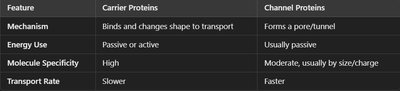
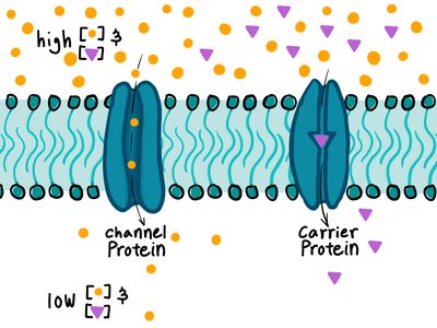
Channel Proteins: Form open pathways for specific ions; transport is usually faster.
Carrier Proteins: Bind specific molecules, change shape to transport; transport is slower and highly specific.
Osmosis and Water Potential
Osmosis is the passive movement of water across a semipermeable membrane, driven by differences in solute concentration.
Hypotonic Solution: Lower solute concentration outside; water enters cell, causing swelling.
Isotonic Solution: Equal solute concentration; no net water movement.
Hypertonic Solution: Higher solute concentration outside; water exits cell, causing shrinkage.
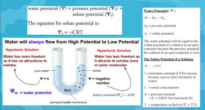
Water Potential Equation:
Solute Potential Equation:
Vocabulary: Lysis (bursting), flaccid (limp), plasmolyzed (shrinkage), turgid (firm).
Active Transport Mechanisms
Types of Active Transport
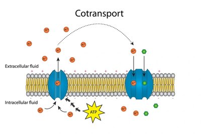
Protein Pumps: Use ATP to move molecules against their concentration gradient (e.g., Na+/K+ pump).
Co-transport: Coupled movement of two molecules; one moves down its gradient, providing energy for the other to move against its gradient.
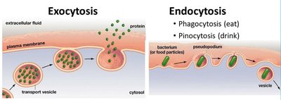
Bulk Transport: Exocytosis and Endocytosis
Exocytosis: Expels materials in vesicles that fuse with the membrane.
Endocytosis: Engulfs materials into vesicles; includes phagocytosis (solids) and pinocytosis (liquids).
Surface Area to Volume Ratio and Membrane Efficiency
Importance of Surface Area
Cells maximize membrane surface area for efficient transport of materials. Structures like microvilli in the small intestine and cristae in mitochondria increase surface area.
Surface Area to Volume Ratio Calculations
As cells grow, volume increases faster than surface area.
Cells function best with a high surface area to volume ratio.
Prokaryotic vs. Eukaryotic Cells
Key Differences
Prokaryotic cells are simple, lacking membrane-bound organelles, while eukaryotic cells are complex and contain organelles. 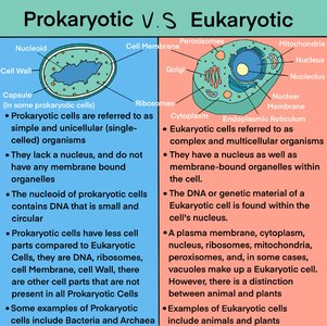
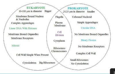
Prokaryotes: Small, no nucleus, circular DNA, no membrane-bound organelles.
Eukaryotes: Larger, nucleus, linear DNA, membrane-bound organelles.
Eukaryotic Organelles: Structure and Function
Overview of Organelles
Eukaryotic cells contain specialized organelles, each with unique structures and functions. 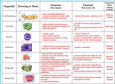
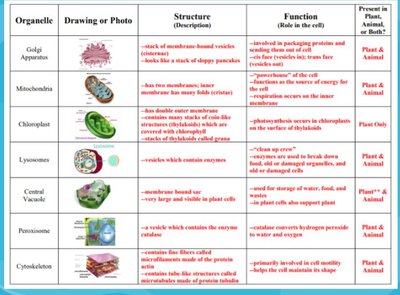
Nucleus: Double membrane, nuclear pores, contains chromatin; regulates gene expression.
Mitochondria: Double membrane, cristae, own DNA; site of ATP production.
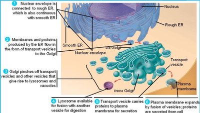
Endoplasmic Reticulum: Rough ER (ribosomes, protein synthesis), Smooth ER (lipid synthesis, detoxification).
Golgi Apparatus: Stacked sacs; modifies, sorts, packages proteins and lipids.
Lysosomes: Vesicles with digestive enzymes; break down waste.
Plasma Membrane: Regulates transport, maintains homeostasis.
Vacuoles: Storage; large central vacuole in plants, small in animals.
Chloroplasts: Photosynthesis in plants; thylakoids and grana.
Cytoskeleton: Microtubules and microfilaments; structure and shape.
Centrosomes/Centrioles: Organize microtubules, involved in mitosis.
Cilia/Flagella: Movement.
Extracellular Matrix: Support and structure.
Intercellular Junctions: Tight junctions, desmosomes, gap junctions, plasmodesmata.
Plant vs. Animal Cells
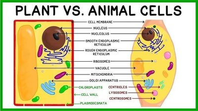
Plant Cells: Cell wall, chloroplasts, large central vacuole, plasmodesmata.
Animal Cells: Centrioles, lysosomes, extracellular matrix.
Endomembrane System and Protein Synthesis
Function and Sequence
The endomembrane system coordinates protein synthesis and secretion.
Proteins are synthesized in the rough ER.
Transported in vesicles to the Golgi apparatus for modification and sorting.
Packaged into vesicles for secretion or delivery to other organelles.
Compartmentalization in Eukaryotic Cells
Role of Membranes
Membrane-bound organelles compartmentalize metabolic processes, minimizing competing interactions and increasing efficiency.
Example: Respiration occurs in mitochondria, transcription/translation in nucleus/ER.
Origins of Eukaryotic Cells: Endosymbiont Theory
Endosymbiont Theory
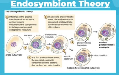
Eukaryotic cells evolved from prokaryotic ancestors through endosymbiosis, where larger cells engulfed smaller ones, forming mitochondria and chloroplasts.
Evidence: Double membranes, own DNA, ribosomes, independent replication, similar size to prokaryotes.
Additional Information
Ribosomes: Made of rRNA and protein; free ribosomes synthesize cytoplasmic proteins, bound ribosomes synthesize membrane/exported proteins.
Water Potential: Measurement of potential energy in water; considers solute and pressure potential.
Cellular Transport: Passive (high to low, no ATP), Active (low to high, ATP).
Osmosis: Water flows from high to low concentration.
Surface Area to Volume Ratio: Cells function best with high ratio.
Major Concepts:
Cell membrane structure and function
Transport mechanisms (passive, active, bulk)
Prokaryotic vs. eukaryotic cell differences
Eukaryotic organelle structure and function
Endomembrane system and compartmentalization
Endosymbiont theory
