 Indietro
Central Dogma, Cell Division, and Genetic Variation: Study Notes for General Biology
Guida di studio - Note intelligenti
 Appunti personalizzati basati sui tuoi materiali, ampliati con definizioni chiave, esempi e contesto.
Central Dogma of Molecular Biology
Overview
The Central Dogma describes the flow of genetic information within a biological system. It explains how DNA is used to create proteins, which are essential for cellular function.
DNA Replication: DNA serves as a template to make more DNA.
Transcription: DNA is transcribed into messenger RNA (mRNA).
Translation: mRNA is translated into a polypeptide (protein).
Key Takeaway: Each step has a distinct purpose: replication for copying DNA, transcription for making RNA, and translation for synthesizing proteins.
DNA Replication
Replication Forks and Bubbles
DNA replication occurs at structures called replication forks and bubbles, where the DNA double helix is unwound and new strands are synthesized.
Leading Strand: Synthesized continuously in the 5' to 3' direction.
Lagging Strand: Synthesized discontinuously as Okazaki fragments.
Key Enzymes:
DNA Polymerase: Synthesizes new DNA strands.
Helicase: Unwinds the DNA helix.
Primase: Adds RNA primers for DNA polymerase to start synthesis.
Ligase: Joins Okazaki fragments together.
Replication forks and bubbles refer to the same concept: regions where DNA is actively being copied. 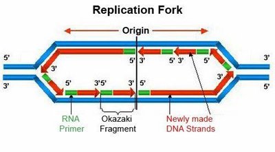
RNA Splicing
Pre-mRNA Processing
Before translation, eukaryotic mRNA undergoes splicing to remove non-coding regions.
Exons: Coding regions that remain in the mature mRNA and exit the nucleus.
Introns: Non-coding regions that are removed (stay in the nucleus).
Splicing: The process of removing introns and joining exons.
Key Point: Only exons are found in mature mRNA; introns are removed. 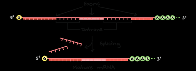
Translation
Protein Synthesis
Translation is the process by which ribosomes synthesize proteins using mRNA as a template.
Direction: mRNA is read from 5' to 3'.
tRNA: Carries amino acids and contains the anticodon that pairs with mRNA codons.
Ribosome Sites:
A site: Entry point for new tRNA.
P site: Holds the tRNA with the growing polypeptide chain.
E site: Exit site for tRNA after amino acid transfer.
Key Point: tRNA is ejected from the E site; anticodon is found on tRNA. 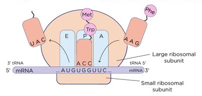
Mitosis
Phases of Mitosis
Mitosis is the process of cell division that produces two genetically identical daughter cells.
Prophase: Chromosomes condense; nuclear membrane breaks down; spindle fibers form.
Metaphase: Chromosomes align at the cell's equator.
Anaphase: Sister chromatids are pulled apart to opposite poles.
Telophase: Nuclear membranes reform; chromosomes decondense.
Cytokinesis: Cell splits into two identical cells.
Key Takeaway: Each step is distinct; do not confuse with meiosis.
Meiosis
Phases and Genetic Variation
Meiosis produces gametes (sperm and egg) with half the chromosome number of the parent cell.
Meiosis I: Separates homologous chromosomes.
Prophase I: Homologs pair and undergo crossing over.
Metaphase I: Homolog pairs align at the center.
Anaphase I: Homologs separate; sister chromatids stay together.
Telophase I + Cytokinesis: Cell splits; chromosomes are still doubled.
Meiosis II: Separates sister chromatids.
Prophase II: Chromosomes condense; spindles form.
Metaphase II: Chromosomes align at the center.
Anaphase II: Sister chromatids separate.
Telophase II + Cytokinesis: Cells split again.
Key Takeaway: Most genetic variation arises in Meiosis I (independent assortment and crossing over).
Cell Cycle
Phases and Functions
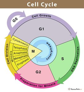
The cell cycle describes the sequence of events in cell growth and division.
G1 Phase: Cell growth and preparation for DNA replication.
S Phase: DNA replication (chromosomes are duplicated).
G2 Phase: Preparation for mitosis.
M Phase: Cell division (mitosis and cytokinesis).
G0 Phase: Resting phase; cells may exit the cycle.
Genetic Variation and Mendelian Genetics
Sources of Genetic Variation
Genetic variation is essential for evolution and is generated through several mechanisms:
Mutation: Changes in DNA sequence.
Independent Assortment: Random alignment of homologous chromosomes during metaphase I of meiosis.
Crossing Over: Exchange of DNA between homologous chromosomes during prophase I.
Random Fertilization: Random combination of gametes.
Meiosis is the key process for genetic variation, especially during Meiosis I.
Mendel's Laws
Law of Independent Assortment: Homologous chromosomes align randomly during metaphase I.
Law of Segregation: Homologous chromosomes separate during anaphase I.
Probability and Test Crosses
Genetic Probability
Probability calculations are used to predict the likelihood of offspring inheriting certain traits.
Test Cross: Used to determine genotype by crossing with a homozygous recessive individual.
Punnett Square: Visual tool for predicting genetic outcomes.
Example: If two carriers (heterozygotes) have a child, the chance of the child being affected by a recessive disease is 25%. The probability for two children both being affected is (6.25%).
Biomolecule Application in Central Dogma
Key Biomolecules
Promoter Sequences: Made of DNA (nucleic acids).
Transcription Factors: Made of proteins.
DNA Transcription
Template and Coding Strands
During transcription, the template strand of DNA is used to synthesize mRNA.
Direction: Template strand runs 3' to 5' from the promoter region.
Transcription Factors: Bind at the promoter region to initiate transcription.
Key Point: The bottom strand (3' to 5' from promoter) is the template strand.
Summary Table: Mitosis vs. Meiosis
Comparison of Cell Division Types
Feature | Mitosis | Meiosis |
|---|---|---|
Number of Divisions | 1 | 2 |
Number of Daughter Cells | 2 | 4 |
Genetic Identity | Identical | Unique |
Chromosome Number | Diploid | Haploid |
Role | Growth, repair | Gamete production |
Practice and Exam Preparation
Key Skills
Fill in codon tables and interpret mRNA sequences.
Use Punnett squares and pedigree tables for genetic analysis.
Draw diagrams illustrating independent assortment and ploidy changes in mitosis vs. meiosis.
Additional info: Practice is essential for mastering these concepts, as exam questions often focus on application and interpretation.
