 Indietro
Extracellular Components and Cell Connections: Coordination of Cellular Activities
Guida di studio - Note intelligenti
 Appunti personalizzati basati sui tuoi materiali, ampliati con definizioni chiave, esempi e contesto.
Extracellular Components and Connections Between Cells
Overview
Cells possess specialized extracellular structures and intercellular connections that are essential for maintaining tissue integrity, communication, and coordination of cellular activities. These features differ between plant and animal cells, reflecting their unique structural and functional requirements.
Cell Walls of Plants
Structure and Function
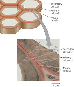
The cell wall is a defining extracellular structure of plant cells, providing protection, shape, and preventing excessive water uptake. It is much thicker than the plasma membrane and is composed of cellulose microfibrils embedded in a matrix of polysaccharides and proteins.
Primary Cell Wall: Thin and flexible, secreted by young plant cells.
Middle Lamella: Rich in pectins, glues adjacent cells together.
Secondary Cell Wall: Added by mature cells, strong and durable, provides extra protection and support (e.g., wood).
Cellulose Synthase: Enzyme responsible for synthesizing cellulose microfibrils.
Example: The architectural design of the cell wall is similar to steel-reinforced concrete, with strong fibers embedded in a matrix.
The Extracellular Matrix (ECM) of Animal Cells
Composition and Function
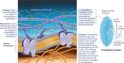
Animal cells lack cell walls but possess an elaborate extracellular matrix (ECM) composed mainly of glycoproteins and carbohydrate-containing molecules. The ECM provides structural support, regulates cell behavior, and facilitates communication between cells.
Collagen: Most abundant glycoprotein, forms strong fibers (about 40% of human body protein).
Proteoglycans: Molecules with a core protein and many carbohydrate chains, forming large complexes.
Fibronectin: Glycoprotein that attaches cells to the ECM.
Integrins: Cell-surface receptor proteins that connect the ECM to the cytoskeleton, transmitting signals and integrating changes inside and outside the cell.
Example: The ECM can influence gene activity in the nucleus through mechanical and chemical signaling pathways.
Cell Junctions
Overview
Cell junctions are specialized structures that facilitate adhesion, communication, and interaction between neighboring cells. They are crucial for the organization of tissues, organs, and organ systems.
Plasmodesmata in Plant Cells
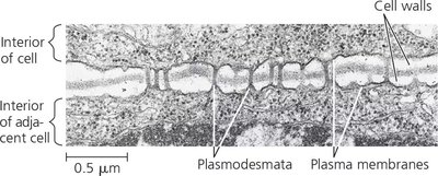
Plasmodesmata are channels that perforate plant cell walls, connecting adjacent cells and allowing the passage of water, solutes, proteins, and RNA. The plasma membranes of adjacent cells are continuous through these channels, unifying most of the plant into one living continuum.
Function: Enables sharing of internal chemical environment and transport of macromolecules.
Structure: Channels lined by plasma membrane, filled with cytosol.
Tight Junctions, Desmosomes, and Gap Junctions in Animal Cells
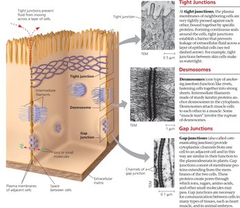
Animal cells possess three main types of cell junctions, especially common in epithelial tissues:
Tight Junctions: Prevent fluid from moving across a layer of cells by tightly pressing membranes together.
Desmosomes: Act like rivets, anchoring junctions that fasten cells together into strong sheets. Intermediate filaments reinforce desmosomes.
Gap Junctions: Provide cytoplasmic channels between adjacent cells, allowing ions, sugars, and other small molecules to pass. Structurally similar to plasmodesmata but formed by proteins rather than membrane.
Example: Gap junctions are essential for communication in cardiac muscle and other tissues requiring rapid signal transmission.
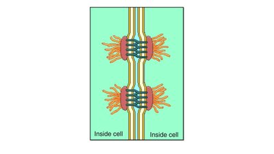
Summary Table: Comparison of Extracellular Structures and Junctions
Structure | Plant Cells | Animal Cells |
|---|---|---|
Extracellular Structure | Cell Wall (cellulose, pectin, etc.) | Extracellular Matrix (collagen, proteoglycans, fibronectin) |
Intercellular Junctions | Plasmodesmata | Tight Junctions, Desmosomes, Gap Junctions |
Function | Protection, shape, water regulation, communication | Support, signaling, adhesion, communication |
Concept Check
Structural Differences: Plant and animal cells have cell walls and ECM, respectively, unlike single-celled eukaryotes.
Permeability: If the cell wall or ECM were impermeable, essential transport and communication would be disrupted, affecting cell function.
Tight Junction Proteins: The amino acid sequence would likely include hydrophobic regions for membrane-spanning segments and hydrophilic regions for loops exposed to the cytoplasm and extracellular space.
Additional info: The notes expand on the brief points in the original material, providing definitions, examples, and a comparison table for clarity and completeness.
