 Indietro
Glial Cells of the Nervous System: Structure, Function, and Development
Guida di studio - Note intelligenti
 Appunti personalizzati basati sui tuoi materiali, ampliati con definizioni chiave, esempi e contesto.
Glial Cells in the Nervous System
Overview of Glial Cell Types
Glial cells, also known as neuroglia, are essential non-neuronal cells in the nervous system that provide structural, metabolic, and functional support to neurons. They are classified based on their location and function in the central nervous system (CNS) and peripheral nervous system (PNS).
Macroglia: Includes astrocytes, oligodendrocytes, ependymal cells, and radial glia in the CNS; Schwann cells and satellite cells in the PNS.
Microglia: Immune cells of the CNS, distinct from macroglia.
Non-glial cells: Vascular endothelial cells and pericytes (CNS).
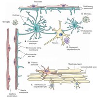
Developmental Origins of Glial Cells
Glial cells arise from different embryonic sources:
Macroglia: Derived from the ectoderm (neural tube or neural crest).
Ependymal cells, endothelial cells, pericytes: Some derived from mesoderm.
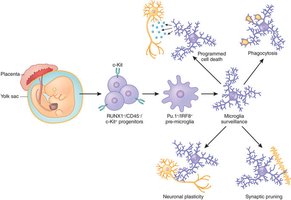
Microglia: Originate from the embryonic yolk sac, similar to macrophages.
Microglia: Immune Cells of the CNS
Microglia Functions and Development
Microglia are the resident immune cells of the CNS, acting as macrophages and antigen-presenting cells. They are crucial for immune defense, surveillance, and maintenance of neural health.
Immune Defense: Constantly survey the brain for pathogens and damage.
Phagocytosis: Engulf and digest dead cells, debris, and toxic aggregates.
Inflammatory Regulation: Release cytokines and chemokines in response to injury.
Synaptic Pruning: Remove unnecessary synapses during development and remodeling.
Synaptic Plasticity: Contribute to learning and memory by modulating synapses.
Support and Repair: Release neurotrophic factors and interact with oligodendrocytes.
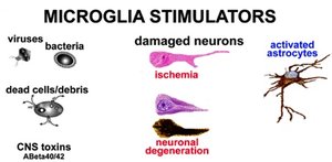
Microglia Activation and Stimulators
Microglia are activated by various stimuli, including pathogens, dead cells, toxins, and damaged neurons.
Stimulators: Viruses, bacteria, dead cells, CNS toxins, ischemia, neuronal degeneration, and activated astrocytes.
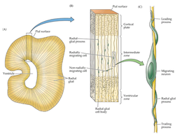
Radial Glia and Development
Radial Glia in the Cerebral Cortex
Radial glial cells guide early neurons to their correct layers during brain development. Initially, they express the astrocyte-specific gene GFAP and later differentiate into cortical neurons or astrocytes.
Function: Scaffold for neuronal migration; precursor to astrocytes.
Ependymal Cells and the Choroid Plexus
Ependymal Cells: Structure and Function
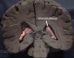
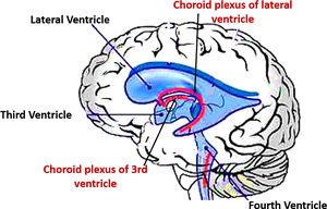
Ependymal cells line the ventricles of the brain and form part of the choroid plexus, which synthesizes cerebrospinal fluid (CSF).
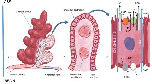
Function: Transport ions, nutrients, and water from capillaries into ventricles; synthesize CSF.
Astrocytes: Structure and Functions
Types of Astrocytes
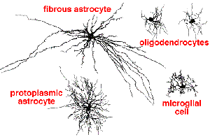
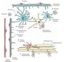
Astrocytes are star-shaped glial cells found in the CNS.
Protoplasmic Astrocytes: Short, thick, highly branched processes; found in gray matter.
Fibrous Astrocytes: Long, thin, less branched processes; found in white matter.
Both types express GFAP, but in different patterns.
Astrocyte Functions
Astrocytes perform multiple roles in the CNS:
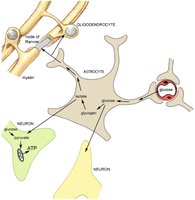
Support neuronal survival: Provide metabolic and trophic support.
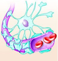
Regulation of capillary diameter: Control blood flow via neurovascular coupling.
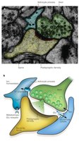
Synaptic development: Regulate synapse formation and stability.
Transport energy substrates: Move glucose and amino acids from blood to neurons and glia.
Regulate extracellular environment: Maintain ion balance and remove excess neurotransmitters.
Tripartite synapse: Participate in synaptic regulation alongside neurons.
Reactive gliosis: Respond to injury by changing phenotype and forming glial scars.
Sources of Adult Astrocytes
Radial glia transformation: Early postnatal development.
Local proliferation: Symmetrical division for maintenance and repair.
Neural stem/progenitor cells: Specialized astrocytes act as stem cells in neurogenic regions.
Oligodendrocytes: Myelinating Cells of the CNS
Oligodendrocyte Structure and Function
Oligodendrocytes form the myelin membrane around axons in the CNS, enabling fast and efficient action potential conduction.
Myelin production: Insulates axons for saltatory conduction.
Metabolic support: Provides nutrients and trophic factors to neurons.
Multiple connections: One oligodendrocyte can myelinate up to 50 axons.
Oligodendrocyte Progenitor Cells (OPCs)
OPCs: Persist throughout the CNS, making up about 5% of neural cells.
NG2-glia: Adult OPCs with potential for differentiation.
Schwann Cells: Myelinating Cells of the PNS
Schwann Cell Functions
Schwann cells myelinate axons in the PNS and support nerve regeneration.
Myelination: Each Schwann cell myelinates a single axon segment.
Support and nutrition: Maintain the chemical environment and nourish neurons.
Unmyelinated support: Form Remak bundles for small axons.
Nerve regeneration: Guide regrowing axons after injury.
Satellite Cells in the PNS
Satellite Cell Functions
Satellite cells surround neuronal cell bodies in ganglia and regulate the local environment, similar to astrocytes in the CNS.
Regulation: Control O2, CO2, nutrients, and neurotransmitters.
Glial Response to Injury
Reactive Gliosis and Regeneration
Reactive gliosis: Astrocytes become reactive, release cytokines, and form glial scars after CNS injury.
CNS regeneration: Damaged axons in the CNS cannot regenerate due to glial scar formation and inhibitory factors.
PNS regeneration: Schwann cells promote axonal regeneration; damaged axons in the PNS can regenerate.
Summary Table: Glial Cell Types and Functions
Cell Type | Location | Main Functions |
|---|---|---|
Astrocyte | CNS | Support, metabolic regulation, synaptic modulation, neurovascular coupling |
Oligodendrocyte | CNS | Myelination, metabolic support |
Microglia | CNS | Immune defense, phagocytosis, synaptic pruning |
Ependymal cell | CNS | CSF synthesis, lining ventricles |
Schwann cell | PNS | Myelination, support, regeneration |
Satellite cell | PNS | Regulation of environment in ganglia |
Key Terms and Concepts
Neuroglia: Non-neuronal support cells in the nervous system.
Myelination: Formation of insulating myelin sheath around axons.
Phagocytosis: Cellular process of engulfing and digesting debris.
Neurovascular coupling: Regulation of blood flow by astrocytes.
Tripartite synapse: Synaptic structure involving neuron-neuron and astrocyte interactions.
Reactive gliosis: Astrocyte response to injury, leading to glial scar formation.
Relevant Equations
Saltatory Conduction Speed: where is conduction velocity, is axon diameter, is membrane resistance, and is membrane capacitance.
Example: Glial Cell Response to Injury
After a stroke, astrocytes become reactive, release inflammatory mediators, and form a glial scar that inhibits axonal regeneration.
In the PNS, Schwann cells clear debris and guide regrowing axons, enabling functional recovery.
Additional info:
Some content was inferred for completeness, such as the saltatory conduction equation and detailed descriptions of glial cell functions.
