 Indietro
Immunological Reactions: Antibodies and Monoclonal Antibodies
Guida di studio - Note intelligenti
 Appunti personalizzati basati sui tuoi materiali, ampliati con definizioni chiave, esempi e contesto.
BASIC PRINCIPLES OF IMMUNOLOGICAL REACTIONS
Introduction to Antibodies
Antibodies, also known as immunoglobulins (Igs), are specialized proteins produced by activated B cells or plasma cells in response to the presence of an antigen. They possess specificity for an epitope of the antigen and play a crucial role in the immune response by binding specifically to antigens.
Definition: Antibodies are proteins that recognize and bind to specific antigens.
Function: They tag antigens for destruction by other immune cells.
Classes: There are five main classes of antibodies, each with distinct structure and function.

Basic Antibody Structure
Polypeptide Composition
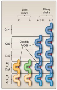
The basic structure of an antibody consists of four polypeptide chains: two identical light chains and two identical heavy chains, linked by disulfide bonds to form a monomeric unit. The chains are aligned so that the amino (NH) terminus of a single heavy and a single light chain form an epitope-binding site.
Variable Domains: Each chain contains a variable domain and one or more constant domains.
Epitope Binding: The variable regions of the light and heavy chains together form the antigen-binding site.

Light Chains
Immunoglobulin monomers contain either two identical κ (kappa) or two identical λ (lambda) light chains, but never one of each. Light chains have a variable (VL) and a constant (CL) domain, each with about 110 amino acids and an intrachain disulfide bond.
Variable Region: Variation in amino acid sequences between immunoglobulins synthesized by different B cells.
Constant Region: Provides structural stability.
Heavy Chains
Heavy chains contain one variable (VH) and three or four constant (CH) domains. There are five isotypes: μ, δ, γ, ε, α. The δ, γ, and α heavy chains have three constant domains, while μ and ε have four, making them longer and heavier.
Isotypes: μ (IgM), δ (IgD), γ (IgG), ε (IgE), α (IgA).
Function: Isotype determines antibody class and function.

Antigen-Binding Sites
The antigen-binding site is formed by the variable domains of both the light and heavy chains, creating a pocket for epitope binding. Each monomeric immunoglobulin has two identical binding sites.
Diversity: Variability in VL and VH domains, along with random pairing, creates a vast pool of binding sites.
Recognition: Enables recognition of numerous different epitopes.
Fragmentation of Antibodies
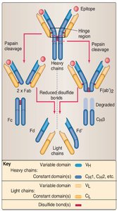
Immunoglobulin molecules can be enzymatically cleaved into discrete fragments by pepsin or papain:
Fab: Antigen-binding fragment
Fc: Constant (crystallizable) fragment
F(ab')2: Dimeric molecule containing two (VH, CH1') segments joined by disulfide bonds
Fd: Heavy chain (VH, CH1) portion of Fab
Fd': Heavy chain (VH, CH1) portion of Fab with extra amino acid

Classes of Antibodies
Overview of Antibody Classes
There are five different classes of antibodies: IgG, IgA, IgM, IgE, and IgD. These classes are defined by the Fc regions in their heavy chains (μ, δ, γ, ε, α) and share the same basic Y-shaped structure.
IgG: Most abundant in serum, activates complement, opsonizes pathogens.
IgA: Found in mucosal secretions, prevents pathogen entry.
IgM: First antibody formed after antigenic stimulation, activates complement.
IgE: Involved in allergic responses, binds to mast cells and basophils.
IgD: Function largely unknown, found on B-cell surfaces.
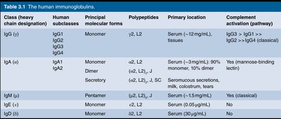
Table: The Human Immunoglobulins
The following table summarizes the main properties of each antibody class:
Class (heavy chain designation) | Human subclasses | Principal molecular forms | Polypeptides | Primary location | Complement activation (pathway) |
|---|---|---|---|---|---|
IgG (γ) | IgG1, IgG2, IgG3, IgG4 | Monomer | γ2, L2 | Serum (~12 mg/mL), tissues | IgG3 > IgG1 > IgG2 (classical) |
IgA (α) | IgA1, IgA2 | Monomer, Dimer, Secretory | α2, L2; (α2, L2, J) | Serum (~3 mg/mL); 90% monomer, 10% dimer; secretory mucous, milk, colostrum, tears | Yes (mannose-binding lectin) |
IgM (μ) | None | Pentamer | μ2, L2, J | Serum (~1.5 mg/mL) | Yes (classical) |
IgE (ε) | None | Monomer | ε2, L2 | Serum (0.05 μg/mL) | No |
IgD (δ) | None | Monomer | δ2, L2 | Serum (30 μg/mL) | No |

IgM
IgM is found as a cell surface-bound monomer or as a secreted pentamer. It is the first immunoglobulin formed after antigenic stimulation and is effective at agglutinating antigens and activating the classical complement pathway.
Structure: Pentameric form in serum.
Function: Agglutination and complement activation.
IgG
IgG is the most abundant immunoglobulin in serum and exists as both surface and secreted monomeric molecules. It has four subclasses and is effective in complement activation, opsonization, neutralization, and antibody-dependent cell-mediated cytotoxicity.
Function: Wide variety of hypersensitivity functions.
Location: Serum and tissues.

IgA
IgA is present in both monomeric (serum) and dimeric forms (mucus, saliva, tears, breast milk, gastrointestinal secretions). It plays a major role in preventing pathogens from entering the body.
Location: Mucosal surfaces, secretory epithelia.
Function: Immune defense at mucosal surfaces.
IgE
IgE is present in low serum concentrations and is mostly adsorbed on the surfaces of mast cells, basophils, and eosinophils. Cross-linking of IgE on mast cell surfaces by antigen triggers the release of histamine and other inflammatory mediators, leading to immediate hypersensitivity (allergic) responses.
Function: Allergic responses and defense against parasites.

IgD
IgD has a monomeric structure and is almost exclusively displayed on B-cell surfaces. Its function is largely unknown.
Location: B-cell surfaces.
Function: Unknown.

Antibody Targets & Functions
Mechanisms of Antibody Action
Antibodies do not directly destroy antigens but inactivate them and tag them for destruction. The formation of antigen-antibody complexes is central to their defensive mechanisms.
Neutralization: Blocking harmful effects of antigens.
Agglutination: Clumping antigens together.
Opsonization: Marking antigens for phagocytosis.
Complement Activation: Triggering complement cascade.
ADCC: Antibody-dependent cell-mediated cytotoxicity.
Monoclonal Antibodies (mAbs)
Production and Properties
Monoclonal antibodies are produced using technology that allows for large quantities of antibodies against specific epitopes. They are made by fusing B lymphocytes with tumor cells, resulting in hybridomas that are long-term sources of pure, identical antibodies.
Hybridomas: Cell hybrids that proliferate endlessly and produce monoclonal antibodies.
Clonal Expansion: Ensures specificity and uniformity of antibodies.
Clinical Uses of Monoclonal Antibodies
Monoclonal antibodies are used in both diagnosis and treatment:
Diagnosis: Pregnancy, allergies, cancer, hepatitis, rabies, and sexually transmitted diseases.
Treatment: Targeted delivery of drugs to cancerous tissue, treatment of autoimmune diseases, leukemia, and lymphomas.
Example: Adalimumab (Humira®) is a recombinant fully human mAb that binds to TNF and neutralizes its function, used in rheumatoid arthritis and Crohn’s disease.
Summary
Antibodies: Y-shaped structure, five classes, distinct functions.
Monoclonal Antibodies: Definition, production, and clinical uses.
