 Indietro
Inside the Cell: Structure and Function of Prokaryotic and Eukaryotic Cells
Guida di studio - Note intelligenti
 Appunti personalizzati basati sui tuoi materiali, ampliati con definizioni chiave, esempi e contesto.
Inside the Cell
Introduction
Cells are the fundamental units of life, and their internal structures collaborate to support the complex processes necessary for survival. This chapter explores the organization, components, and functions of prokaryotic and eukaryotic cells, as well as the dynamic systems that maintain cellular function.
Prokaryotic Cell Structures and Their Functions
Overview of Prokaryotic Cells
Prokaryotic cells, which include Bacteria and Archaea, lack a membrane-bound nucleus but possess a variety of specialized structures:
Chromosome: Usually a single, circular DNA molecule associated with proteins, located in a region called the nucleoid.
Plasmids: Small, circular DNA molecules that carry genes beneficial for survival.
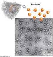
Ribosomes: Macromolecular complexes of RNA and protein responsible for protein synthesis.
Cytoplasm: The internal fluid containing all cellular contents.
Cell Wall: Provides structural support and shape; in bacteria, primarily composed of peptidoglycan.
Plasma Membrane: A selectively permeable barrier composed of phospholipids (fatty acids in Bacteria, branched isoprenoids in Archaea).
Cytoskeleton: Protein filaments that maintain cell shape and assist in division.
Photosynthetic Membranes: Present in some species, these internal membranes contain pigments and enzymes for photosynthesis.
Organelles: Some prokaryotes have membrane-bound compartments for specialized functions (e.g., storing ions, orienting with magnetite crystals).
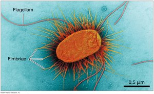
External Structures: Flagella (for movement) and fimbriae (for attachment).
Eukaryotic Cell Structures and Their Functions
General Features
Eukaryotic cells are typically larger and more complex than prokaryotic cells. They contain numerous membrane-bound organelles that compartmentalize cellular functions.
Nucleus: Contains genetic material (DNA), surrounded by a double-membrane nuclear envelope with nuclear pores for selective transport. The nucleolus within is the site of ribosomal RNA synthesis and ribosome assembly.
Ribosomes: Sites of protein synthesis, found free in the cytosol or bound to the endoplasmic reticulum (ER).
Endoplasmic Reticulum (ER):
Rough ER (RER): Studded with ribosomes; synthesizes and processes proteins for secretion or membrane insertion.
Smooth ER (SER): Lacks ribosomes; involved in lipid synthesis and detoxification.
Golgi Apparatus: Stacks of cisternae that process, sort, and ship proteins and lipids received from the ER.
Lysosomes: Contain hydrolytic enzymes for digestion and recycling of macromolecules (mainly in animal cells).
Vacuoles: Large storage organelles in plants and fungi, involved in storage, digestion, and maintaining turgor pressure.
Peroxisomes: Sites of oxidation-reduction reactions; detoxify harmful substances (e.g., hydrogen peroxide).
Mitochondria: Powerhouses of the cell, generating ATP through cellular respiration; contain their own DNA and ribosomes.
Chloroplasts: Found in plants and algae; sites of photosynthesis, also containing their own DNA and ribosomes.
Cytoskeleton: Network of protein fibers (actin filaments, intermediate filaments, microtubules) providing structure, transport, and movement.
Cell Wall: Present in plants, fungi, and algae; provides structural support and protection.
How the Parts Fit into a Whole
Structure-Function Relationships
The structure of each cell component is closely related to its function. For example, fat cells are specialized for lipid storage, while muscle cells are elongated for contraction. The number and type of organelles vary according to the cell’s role.
Nuclear Transport
Selective Transport through the Nuclear Envelope
The nuclear envelope separates the nucleus from the cytoplasm and contains nuclear pore complexes that regulate the movement of molecules. Proteins destined for the nucleus contain a nuclear localization signal (NLS), a specific amino acid sequence that acts as a molecular "zip code" for import.
The Endomembrane System: Manufacturing, Shipping, and Recycling
Secretory Pathway and Protein Targeting
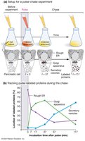
The endomembrane system includes the ER, Golgi apparatus, lysosomes, and vesicles. Proteins synthesized in the RER are transported to the Golgi apparatus for further processing and sorting. The pulse–chase experiment demonstrated the movement of proteins through this pathway.
Signal Hypothesis
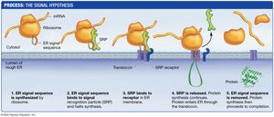
Proteins destined for the endomembrane system contain an ER signal sequence that directs them to the RER. The signal recognition particle (SRP) binds the sequence and guides the ribosome to the ER membrane, where the protein is synthesized into the lumen and processed (e.g., glycosylation).
Protein Sorting and Vesicle Transport
Proteins are tagged with molecular markers in the Golgi apparatus, ensuring delivery to the correct destination (e.g., lysosomes, plasma membrane). Transport vesicles bud from the Golgi and fuse with target membranes, releasing their contents.
Lysosomal Recycling Pathways
Receptor-mediated endocytosis: Uptake of specific molecules via receptor binding and vesicle formation.
Phagocytosis: Engulfment of large particles or cells.
Autophagy: Digestion of the cell’s own damaged organelles or macromolecules.
The Dynamic Cytoskeleton
Types of Cytoskeletal Elements
Actin Filaments (Microfilaments): Smallest, composed of actin; involved in cell shape, movement, and division.
Intermediate Filaments: Provide mechanical support; include keratins and nuclear lamins.
Microtubules: Largest, hollow tubes made of tubulin; serve as tracks for vesicle transport and are essential for chromosome separation during cell division.
Motor Proteins and Vesicle Transport
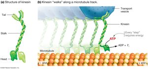
Kinesin is a motor protein that "walks" along microtubules, transporting vesicles by converting ATP into mechanical energy.
Flagella and Cilia
Flagella and cilia are cellular appendages used for movement. Eukaryotic flagella and cilia have a "9 + 2" arrangement of microtubules (axoneme) and move via the action of the motor protein dynein, which causes bending and movement.
Table: Comparison of Prokaryotic and Eukaryotic Cell Structures
Feature | Prokaryotic Cells | Eukaryotic Cells |
|---|---|---|
Nucleus | Absent | Present |
Organelles | Few, not membrane-bound | Numerous, membrane-bound |
Cell Size | Generally smaller | Generally larger |
DNA Structure | Circular, single chromosome | Linear, multiple chromosomes |
Cell Wall | Usually present (peptidoglycan in bacteria) | Present in plants, fungi, algae (cellulose, chitin, etc.) |
Examples | Bacteria, Archaea | Protists, fungi, plants, animals |
Key Terms and Concepts
Organelle: Specialized structure within a cell that performs a specific function.
Endomembrane System: Network of membranes involved in protein and lipid synthesis, modification, and transport.
Glycosylation: Addition of carbohydrate groups to proteins.
Pulse–Chase Experiment: Technique to track the movement of molecules through cells.
Motor Protein: Protein that converts chemical energy into mechanical work (e.g., kinesin, dynein, myosin).
Axoneme: Core structure of cilia and flagella, composed of microtubules in a "9 + 2" arrangement.
