 Indietro
Inside the Cell: Structure and Function of Prokaryotic and Eukaryotic Cells
Guida di studio - Note intelligenti
 Appunti personalizzati basati sui tuoi materiali, ampliati con definizioni chiave, esempi e contesto.
Inside the Cell
The Scale of Life
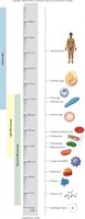
Cells and their components vary greatly in size, from molecules to multicellular organisms. Most cell diameters are in the range of 1–100 μm, which allows for efficient transport and communication within the cell.
Relative Sizes: Molecules < Organelles < Cells < Multicellular Organisms
Microscopy: Essential for visualizing cell structures at these scales.
Prokaryotic Cells
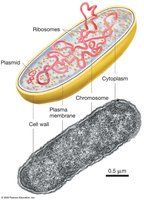
Basic Structure of Prokaryotic Cells
Prokaryotic cells, such as bacteria and archaea, are characterized by their simple structure and lack of membrane-bound organelles.
Cell Wall: Provides structural support and protection.
Plasma Membrane: Encloses the cytoplasm and regulates transport.
Cytoplasm: Contains all cellular components.
Nucleoid: Region where the single, circular chromosome is located.
Ribosomes: Sites of protein synthesis.
Plasmids: Small, circular DNA molecules with extra genetic information.
Prokaryotic DNA Organization
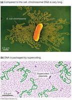
Prokaryotic DNA is often supercoiled to fit within the cell and may include plasmids, which carry genes for specialized functions.
Supercoiling: Compacts DNA for efficient storage.
Plasmids: Can confer advantages such as antibiotic resistance.
Specialized Prokaryotic Organelles
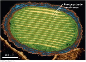
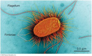
Certain prokaryotes possess internal membranes for photosynthesis and external appendages for movement and attachment.
Photosynthetic Membranes: Increase surface area for light absorption and energy production.
External Appendages: Flagella for movement; fimbriae for attachment.
Eukaryotic Cells
Animal Eukaryotic Cell Structure
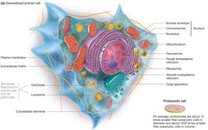
Animal cells are complex, with numerous membrane-bound organelles and specialized structures.
Nucleus: Contains DNA and controls cellular activities.
Mitochondria: Powerhouse of the cell, site of ATP production.
Centrioles: Involved in cell division.
Lysosomes: Recycling centers containing digestive enzymes.
Plant Eukaryotic Cell Structure
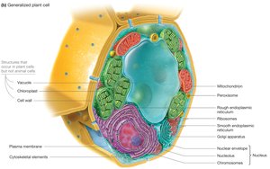
Plant cells share many features with animal cells but also have unique structures.
Cell Wall: Provides rigidity and protection.
Chloroplasts: Site of photosynthesis.
Vacuole: Stores water and nutrients.
DNA Location: Nucleus, mitochondria, and chloroplasts.
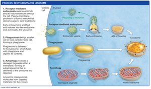
Lysosomes: Recycling Centers
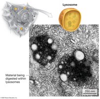
Lysosomes are specialized organelles in animal cells responsible for breaking down macromolecules using acid hydrolases.
Acid Hydrolases: Digestive enzymes active at pH 5.0.
Proton Pumps: Maintain acidic environment inside lysosomes.
Pathways to Lysosome for Recycling
Material is directed to lysosomes via three main pathways: receptor-mediated endocytosis, phagocytosis, and autophagy.
Receptor-Mediated Endocytosis: Uptake of macromolecules.
Phagocytosis: Engulfment of large particles or cells.
Autophagy: Digestion of damaged organelles.
The Endomembrane System
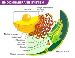
The endomembrane system is a network of organelles that produce, process, and transport proteins, carbohydrates, and lipids in eukaryotic cells.
Components: Endoplasmic reticulum (rough and smooth), Golgi apparatus, lysosomes.
Function: Synthesis, modification, and transport of biomolecules.
Mitochondria: Power Generating Station
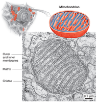
Mitochondria are double-membraned organelles responsible for cellular respiration and ATP production. They contain their own DNA and are found in both animal and plant cells.
Structure: Outer and inner membranes, matrix, cristae.
Function: Oxidize food molecules to produce ATP.
Chloroplasts: Sugar Manufacturing Center
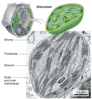
Chloroplasts are double-membraned organelles in plant cells that absorb light and use its energy to produce carbohydrates from CO2. They also contain their own DNA.
Structure: Outer and inner membranes, stroma, thylakoids, granum.
Function: Photosynthesis.
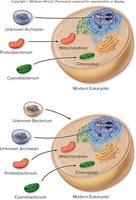
Endosymbiosis Theory
The endosymbiosis theory proposes that mitochondria and chloroplasts originated as free-living bacteria that were engulfed by ancestral eukaryotic cells, leading to a mutually beneficial relationship.
Evidence: Both organelles contain their own DNA and replicate independently.
Implication: Explains the presence of prokaryotic-like features in these organelles.
Cytoskeleton: Non-Membranous Organelles
Types of Cytoskeletal Elements
The cytoskeleton is an extensive system of protein fibers that provides structural support, facilitates movement, and organizes the cell's contents.
Actin Filaments (Microfilaments): Maintain cell shape and enable movement.
Intermediate Filaments: Provide structural support; not involved in movement.
Microtubules: Large protein fibers involved in cell division and transport.
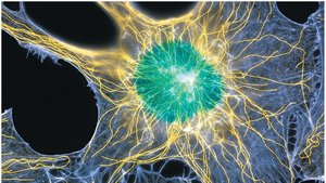
Microtubules
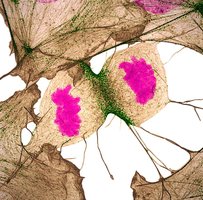
Microtubules are dynamic structures that polymerize and depolymerize, serving as tracks for vesicle transport and playing a crucial role in chromosome separation during cell division.
Centrosome: Microtubule-organizing center in animal cells, containing centrioles.
Vesicle Transport: Motor proteins like kinesin move vesicles along microtubules using ATP.
Actin Filaments
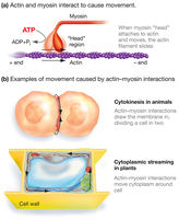
Actin filaments are found just inside the plasma membrane and help define the cell’s shape. They have distinct plus and minus ends and interact with myosin to facilitate movement.
Cell Shape: Maintained by actin filaments.
Movement: Actin-myosin interactions cause muscle contraction, cytokinesis, and cytoplasmic streaming.
Intermediate Filaments
Intermediate filaments provide structural support for the cell and are not involved in movement. Nuclear laminins, a type of intermediate filament, form a flexible skeleton that shapes the cell surface and holds the nucleus in place.
Structural Support: Maintains cell integrity.
Nuclear Laminins: Support the nuclear envelope.
Key Differences Between Eukaryotes and Prokaryotes
Feature | Eukaryotes | Prokaryotes |
|---|---|---|
Nucleus | Present | Absent (Nucleoid) |
Organelles | Present | Absent |
Cell Structure | Larger, compartmentalized | Smaller, less compartmentalized |
Cytoskeleton | Diverse and dynamic | Rudimentary |
Cell Structure Correlates with Function
The abundance of certain organelles in a cell reflects its specialized function. For example, cells packed with rough ER are specialized for protein synthesis, while those with smooth ER are involved in lipid synthesis. Cells rich in mitochondria or chloroplasts are specialized for energy production or photosynthesis, respectively.
Rough ER: Protein synthesis.
Smooth ER: Lipid synthesis.
Mitochondria: ATP production.
Chloroplast: Photosynthesis.
Additional info: The notes above expand on brief points with academic context, definitions, and examples to ensure completeness and clarity for exam preparation.
