 Indietro
CH. 7 - Inside the Cell: Structure and Function of Cellular Components
Guida di studio - Note intelligenti
 Appunti personalizzati basati sui tuoi materiali, ampliati con definizioni chiave, esempi e contesto.
Inside the Cell
Cell Theory and Basic Cell Components
Cell theory states that all living organisms are composed of cells, which are the fundamental units of life. Cells share several universal features that enable their function and survival.
Proteins: Perform most cellular functions, including catalysis, structure, and signaling.
Nucleic acids: Store, transmit, and process genetic information (DNA and RNA).
Carbohydrates: Provide chemical energy, carbon, structural support, and cellular identity.
Plasma membrane: Acts as a selectively permeable barrier, regulating entry and exit of substances.
Cell Types and Domains of Life
Cells are classified based on morphology and evolutionary history:
Eukaryotes: Possess a membrane-bound nucleus.
Prokaryotes: Lack a membrane-bound nucleus.
Three Domains: Bacteria (prokaryotic), Archaea (prokaryotic), Eukarya (eukaryotic).
Prokaryotic Cell Structures
Structural Components of Prokaryotic Cells
Prokaryotic cells exhibit a variety of specialized structures and functions.
Contain at least one chromosome and numerous ribosomes for protein synthesis.
Phospholipid composition: Bacterial phospholipids have fatty acids bound to glycerol; archaeal phospholipids use branched isoprenoid chains.
Cytoplasm: All contents within the cell membrane.
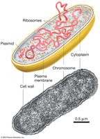
Chromosome Organization and Plasmids
The chromosome is the most prominent structure in prokaryotic cells, typically a single, circular DNA molecule associated with proteins. Plasmids are smaller, circular, supercoiled DNA molecules that carry additional genes.
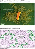
Ribosomes and Protein Synthesis
Prokaryotic ribosomes are macromolecular machines composed of RNA and protein, responsible for synthesizing proteins. Bacterial and archaeal ribosomes are similar in size and function but differ in primary structure.
Cytoskeleton and Cell Shape
Long, thin protein filaments form the cytoskeleton, essential for cell division and maintaining cell shape in prokaryotes.
Internal Membrane Complexes in Photosynthetic Species
Photosynthetic prokaryotes possess internal membrane complexes that convert sunlight into chemical energy. These membranes are infoldings of the plasma membrane and contain enzymes and pigments necessary for photosynthesis.
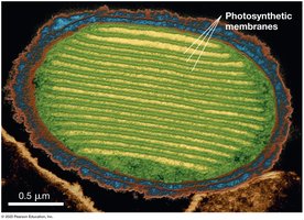
Organelles for Specialized Functions
Some bacteria have internal, membrane-bound compartments (organelles) for specialized tasks, such as storing ions, orienting with magnetite crystals, or concentrating enzymes.
Cell Wall Structure
The cell wall forms a protective exoskeleton, providing shape and rigidity. In bacteria, the primary structural component is peptidoglycan, a polysaccharide. Some bacteria also have an outer membrane made of glycolipids.
External Structures: Flagella and Fimbriae
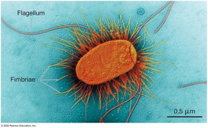
Prokaryotes interact with their environment using external structures:
Flagella: Long filaments that rotate to propel the cell.
Fimbriae: Needlelike projections that promote attachment to surfaces or other cells.
Eukaryotic Cell Structures and Functions
Overview of Eukaryotic Cells
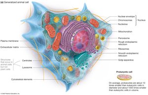
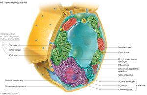
Eukaryotes include protists, fungi, plants, and animals, and may be unicellular or multicellular. Eukaryotic cells are generally larger (5–100 µm) than prokaryotic cells (1–10 µm).
Benefits of Organelles
Organelles compartmentalize the cell, allowing separation of incompatible reactions and increasing efficiency. The cytosol is the fluid portion outside organelles.
The Nucleus
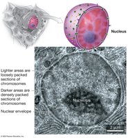
The nucleus is a large, highly organized, membrane-bound compartment. It is surrounded by a double-membrane nuclear envelope with pore-like openings and contains the nucleolus, where ribosomal RNA is synthesized and ribosome subunits are assembled.
Ribosomes
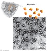
Ribosomes are complex molecular machines that manufacture proteins. They lack a membrane and can be free in the cytosol or attached to the endoplasmic reticulum.
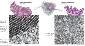
Endoplasmic Reticulum (ER)
The ER is an extensive membrane-enclosed factory, continuous with the nuclear envelope. It has two regions:
Rough ER (RER): Studded with ribosomes, synthesizes proteins for shipment, membrane insertion, or secretion.
Smooth ER (SER): Lacks ribosomes, contains enzymes for lipid synthesis and detoxification, and stores Ca2+ ions.
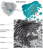
Golgi Apparatus
The Golgi apparatus processes, sorts, and ships proteins synthesized in the RER. It consists of stacked, flat membranous sacs (cisternae) with distinct cis (entry) and trans (exit) faces.
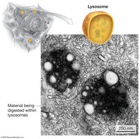
Lysosomes
Lysosomes are recycling centers found only in animal cells, containing about 40 different enzymes (acid hydrolases) that hydrolyze macromolecules. They maintain a low internal pH via proton pumps.
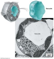
Vacuoles
Vacuoles are prominent organelles in plant and fungal cells, serving as storage centers for water, ions, proteins, pigments, and defensive compounds.
Peroxisomes
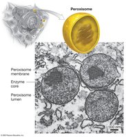
Peroxisomes are globular organelles that perform reduction–oxidation (redox) reactions, often producing and detoxifying hydrogen peroxide.
Mitochondria
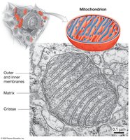
Mitochondria supply ATP to cells and have two membranes. The inner membrane is folded into cristae, enclosing the mitochondrial matrix. Mitochondria have their own DNA and ribosomes, and can grow and divide independently.
Chloroplasts
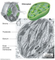
Chloroplasts are found in plant and algal cells, where photosynthesis occurs. They have three membranes, with thylakoids arranged in stacks (grana) and surrounded by stroma. Chloroplasts contain their own DNA and ribosomes.
Cell Wall and Extracellular Matrix
Fungi, algae, and plants have a stiff cell wall outside the plasma membrane for structural support. Animal cells lack a cell wall but are supported by the extracellular matrix (ECM), a mixture of secreted proteins and polysaccharides.
Cell Structure and Function Correlation
Variation in Organelle Content
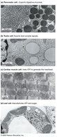
The structure and number of organelles in a cell correlate with its specialized function. For example, fat cells store lipids, cardiac muscle cells generate ATP, and leaf cells contain chloroplasts for photosynthesis.
Nuclear Transport
Nuclear Envelope and Pore Complexes
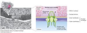
The nuclear envelope separates the nucleus from the cytosol and is perforated with nuclear pore complexes, which regulate the selective import and export of molecules.
Nuclear Localization Signals
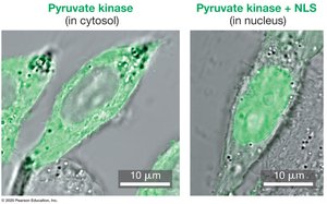
Proteins destined for the nucleus contain a nuclear localization signal (NLS), a 17-amino-acid sequence that acts as a molecular zip code for transport through the nuclear pore complex.
The Endomembrane System
Secretory Pathway Hypothesis
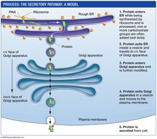
The endomembrane system includes the ER, Golgi apparatus, and lysosomes, and is responsible for producing, processing, and transporting proteins, carbohydrates, and lipids. Proteins are actively imported from the cytosol and require specific zip codes for correct localization.
Pulse–Chase Experiment
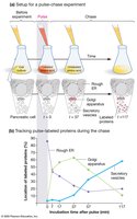
The pulse–chase experiment tracks protein movement through the secretory pathway, demonstrating that proteins move from the RER to the Golgi apparatus and then to secretory vesicles or the cell exterior.
Signal Hypothesis for Protein Targeting
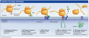
Proteins bound for the endomembrane system have an ER signal sequence, a 20-amino-acid zip code that directs the growing polypeptide to the RER.
Protein Sorting and Vesicle Transport
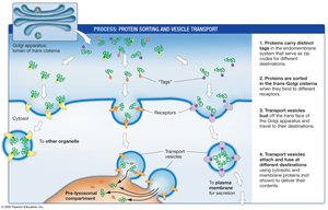
Proteins are sorted in the Golgi apparatus and tagged for delivery to specific destinations. Transport vesicles carry proteins to organelles or the plasma membrane for secretion.
Lysosomal Recycling Pathways
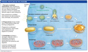
Cells recycle materials via lysosomes through three pathways: receptor-mediated endocytosis, phagocytosis, and autophagy. These processes allow digestion and reuse of macromolecules and damaged organelles.
The Dynamic Cytoskeleton
Cytoskeletal Elements
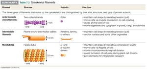
The cytoskeleton is a dynamic network of protein fibers that maintains cell shape, transports materials, and organizes cellular structures. It consists of three major types:
Actin filaments (microfilaments): Smallest, made of actin, involved in movement and cell shape.
Intermediate filaments: Provide structural support, anchor organelles, and stabilize the nucleus.
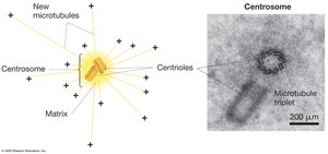
Microtubules: Largest, made of tubulin dimers, involved in chromosome separation, vesicle transport, and cell movement.
Actin Filament Function
Actin filaments interact with the motor protein myosin to produce movement, including cytokinesis, cytoplasmic streaming, and cell crawling.
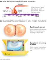
Microtubule Organization and Vesicle Transport
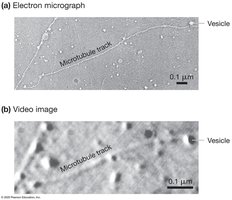
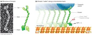
Microtubules originate from the microtubule organizing center (MTOC), called the centrosome in animals. They serve as tracks for vesicle transport, with the motor protein kinesin converting ATP into mechanical energy to "walk" vesicles along microtubules.
Flagella and Cilia
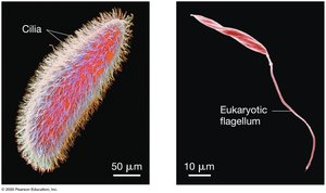
Flagella and cilia are hairlike projections that move cells or substances. Prokaryotic flagella rotate, while eukaryotic flagella and cilia have a "9 + 2" microtubule arrangement and move via bending.
Cell-to-Cell Junctions
Intercellular Junctions in Plants and Animals
Multicellular organisms use cell junctions for adhesion, interaction, and communication:
Plants: Plasmodesmata allow cytoplasmic continuity and transport between cells.
Animals: Tight junctions prevent leakage, desmosomes anchor cells, and gap junctions provide cytoplasmic channels for communication.
Table 7.1 Eukaryotic Cell Components
Organelle | Structure | Function |
|---|---|---|
Nucleus | Double membrane, nuclear pores | Stores genetic information, site of RNA synthesis |
Ribosomes | No membrane, RNA and protein | Protein synthesis |
Rough ER | Membrane with ribosomes | Protein synthesis and processing |
Smooth ER | Membrane without ribosomes | Lipid synthesis and detoxification |
Golgi apparatus | Stacked cisternae | Protein processing, sorting, shipping |
Lysosomes | Single membrane, acid hydrolases | Macromolecule digestion and recycling |
Vacuoles | Large membrane-bound sac | Storage and digestion |
Peroxisomes | Single membrane, enzymes | Redox reactions, detoxification |
Mitochondria | Double membrane, cristae | ATP production |
Chloroplasts | Double membrane, thylakoids | Photosynthesis |
Cell wall | Rigid outer layer | Structural support |
