 Indietro
Polygenic Inheritance, GWAS, and CRISPR Applications in Human Genetics
Guida di studio - Note intelligenti
 Appunti personalizzati basati sui tuoi materiali, ampliati con definizioni chiave, esempi e contesto.
Polygenic Inheritance and Quantitative Traits
Definition and Characteristics
Polygenic inheritance refers to the phenomenon where multiple genes contribute additively to a single phenotype, resulting in continuous variation within a population. Traits governed by polygenic inheritance are called quantitative characters, as they do not fall into discrete categories but instead show a range of phenotypes.
Quantitative traits include height, skin color, and intelligence in humans.
Each gene involved typically has a small effect, but collectively, they produce significant phenotypic diversity.
Polygenic traits often display a normal distribution (bell-shaped curve) in the population.
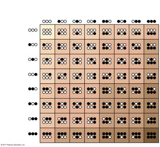
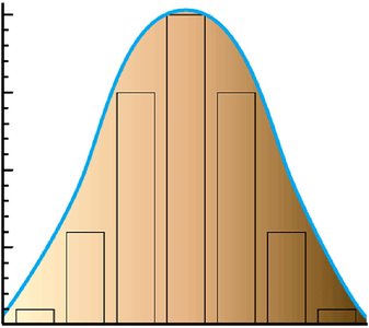
Example: Human skin color is influenced by at least three genes, each with two alleles, resulting in a wide range of possible skin tones.
Genome-Wide Association Studies (GWAS)
Principles and Methodology
GWAS is a powerful approach for identifying genetic variants associated with complex traits and diseases in humans. It relies on analyzing genetic variation across the genome in large populations to find statistical associations between specific genetic markers and phenotypes.
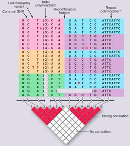
Single nucleotide polymorphisms (SNPs) are the most common type of genetic variation used in GWAS.
GWAS does not require controlled crosses; instead, it leverages natural genetic variation accumulated over generations.
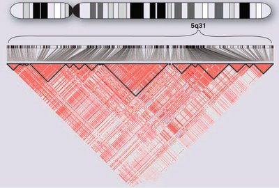
Closely linked polymorphisms are inherited together due to limited recombination, forming haplotype blocks.
Applications and Interpretation
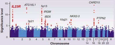
GWAS can identify loci associated with diseases such as Crohn's disease, height, and other polygenic traits.
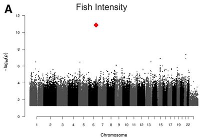
Results are often visualized as Manhattan plots, where each dot represents a SNP and the y-axis indicates the statistical significance of association.
Significant SNPs are not necessarily causative but are genetically linked to the trait.
Large sample sizes and advanced statistical methods are required for robust results.
Example: GWAS identified a missense mutation in the TAAR5 gene associated with human perception of the odor of rotten fish.
Hemoglobin Structure and Genetic Disorders
Hemoglobin Structure and Gene Families
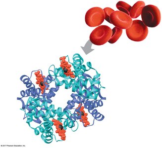
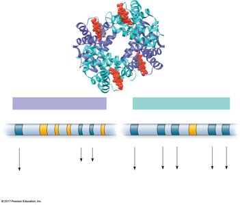
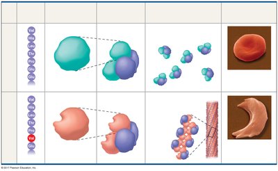
Hemoglobin is a tetrameric protein in red blood cells responsible for oxygen transport. It consists of two alpha and two beta subunits, each encoded by separate gene families located on different chromosomes.
Alpha-globin gene family: Located on chromosome 16.
Beta-globin gene family: Located on chromosome 11.
Different hemoglobin types are expressed during development: embryonic (HbE), fetal (HbF), and adult (HbA).
Sickle-Cell Disease and Beta-Thalassemia
Both disorders are caused by mutations in the beta-globin gene, affecting hemoglobin function and red blood cell morphology.
Sickle-cell disease: Caused by a missense mutation (Glu to Val) in the beta-globin gene, leading to hemoglobin aggregation and sickle-shaped red blood cells.
Beta-thalassemia: Caused by a variety of mutations (e.g., in promoter, splicing, or coding regions) that reduce or eliminate beta-globin production.
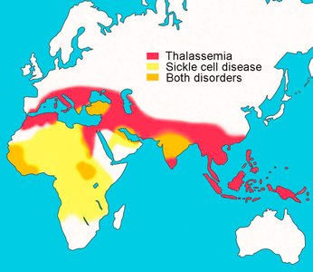
Both diseases are prevalent in regions where malaria is or was common, due to heterozygote advantage.
Hereditary Persistence of Fetal Hemoglobin (HPFH)
Genetic Basis and Clinical Significance
HPFH is a benign condition where individuals continue to express high levels of fetal hemoglobin (HbF) into adulthood, which can ameliorate symptoms of sickle-cell disease and beta-thalassemia.
HPFH can result from deletions in the beta-globin gene cluster or point mutations in the gamma-globin gene promoter.
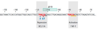
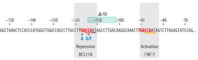
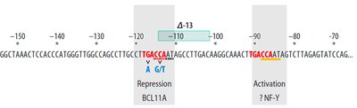
Mutations in a control element (TGACCA motif) in the proximal promoter of the gamma-globin gene disrupt binding of the BCL11A transcription factor, relieving repression and increasing HbF expression.
GWAS and Identification of Regulatory Loci
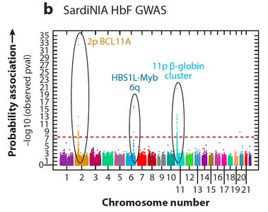
GWAS identified several loci associated with HPFH, including the BCL11A gene on chromosome 2, which encodes a transcription factor that represses gamma-globin expression.
BCL11A binds to the TGACCA motif in the gamma-globin promoter, acting as a repressor.
SNPs in intron 2 of BCL11A overlap with enhancer elements, affecting BCL11A expression and thus HbF levels.
CRISPR-Based Therapeutic Strategies
Gene Editing to Treat Hemoglobinopathies
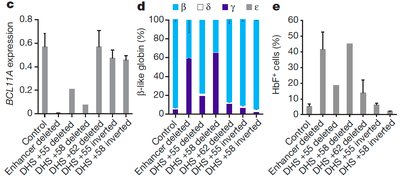
CRISPR-Cas9 technology enables targeted editing of the genome to disrupt regulatory elements and relieve repression of beneficial genes. In the context of sickle-cell disease and beta-thalassemia, CRISPR can be used to knock out the BCL11A enhancer, increasing HbF production and alleviating disease symptoms.
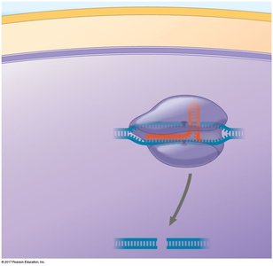
CRISPR guide RNA targets the enhancer in intron 2 of BCL11A, causing a double-strand break.
Repair by non-homologous end joining disrupts the enhancer, reducing BCL11A expression and increasing gamma-globin (HbF) expression.
Clinical Application and Outcomes
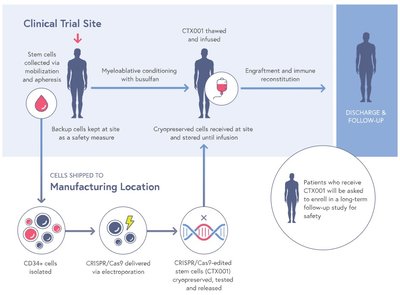
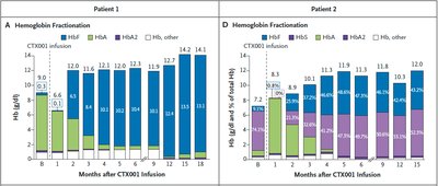
In clinical trials, patient stem cells are edited ex vivo using CRISPR, then reinfused after myeloablative conditioning. This approach has led to increased HbF levels and clinical improvement in patients with sickle-cell disease and beta-thalassemia.
Patients show sustained increases in HbF and reduction or elimination of disease symptoms.
Long-term follow-up is ongoing to assess safety and efficacy.
Summary Table: Comparison of Hemoglobin Disorders and Treatments
Disorder | Genetic Cause | Effect on Hemoglobin | Therapeutic Approach |
|---|---|---|---|
Sickle-cell disease | Missense mutation (Glu→Val) in β-globin | Hemoglobin S aggregates, sickled RBCs | Gene addition, gene correction, HbF induction (CRISPR) |
β-thalassemia | Various mutations in β-globin gene | Reduced/absent β-globin, anemia | Gene addition, gene correction, HbF induction (CRISPR) |
HPFH | Deletions or promoter mutations in β-globin cluster | Persistent fetal hemoglobin expression | Benign; leveraged for therapy |
Key Concepts Review
Polygenic inheritance underlies most human traits, resulting in quantitative variation.
GWAS enables identification of genetic loci associated with complex traits and diseases.
Hemoglobinopathies such as sickle-cell disease and β-thalassemia are caused by mutations in the β-globin gene.
HPFH provides a natural model for therapeutic strategies that increase fetal hemoglobin expression.
CRISPR-Cas9 gene editing offers a promising approach to treating genetic diseases by targeting regulatory elements.
