 Indietro
Structure and Function of DNA: Study Notes for General Biology
Guida di studio - Note intelligenti
 Appunti personalizzati basati sui tuoi materiali, ampliati con definizioni chiave, esempi e contesto.
Structure and Function of DNA
Discovering the Structure of DNA
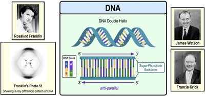
The discovery of DNA's structure was a pivotal moment in biology, revealing the molecular basis of heredity. In the early 1950s, Rosalind Franklin used X-ray diffraction to capture images of DNA, most notably "Photo 51," which provided critical evidence for the double helix structure. James Watson and Francis Crick used Franklin's data to describe DNA as a double helix with two anti-parallel strands of nucleotides. The base-pairing rules, established by Watson and Crick, state that nucleotides on opposite strands pair via hydrogen bonds: Adenine (A) pairs with Thymine (T), and Cytosine (C) pairs with Guanine (G).
Double Helix: DNA consists of two strands twisted into a helical shape.
Anti-parallel Strands: The two strands run in opposite directions (5' to 3' and 3' to 5').
Base Pairing: A–T and C–G pairs are held together by hydrogen bonds.
Key Scientists: Franklin (X-ray diffraction), Watson & Crick (structure), Chargaff (base ratios).
Detailed DNA Structure
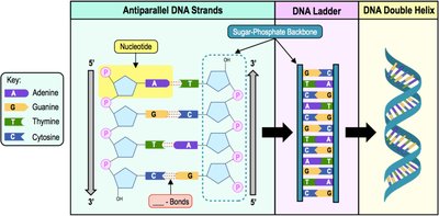
DNA is a polymer made of nucleotide monomers, each consisting of a phosphate group, a deoxyribose sugar, and a nitrogenous base. The strands are linked by phosphodiester bonds, forming a sugar-phosphate backbone. Each strand has a free phosphate group at the 5' end and a free hydroxyl group (-OH) at the 3' end. The anti-parallel arrangement and complementary base pairing are essential for DNA replication and function.
Nucleotide: Basic unit of DNA, composed of phosphate, sugar, and base.
Phosphodiester Bond: Links the 5' phosphate of one nucleotide to the 3' OH of the next.
Hydrogen Bonds: Hold the two strands together between bases.
DNA Replication
Introduction to DNA Replication
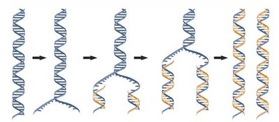
DNA replication is the process by which a cell copies its DNA before cell division. The parental strands separate and serve as templates for the synthesis of new complementary strands. This process is fundamental in both prokaryotes and eukaryotes, though more is known about prokaryotic replication.
Template Strands: Parental DNA strands guide the synthesis of new DNA.
Complementary Base Pairing: Ensures accurate copying of genetic information.
Components of DNA Replication
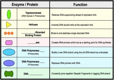
DNA replication requires several enzymes and proteins working together:
Enzyme / Protein | Function |
|---|---|
Topoisomerase | Relieves DNA supercoiling ahead of replication fork |
Helicase | Unwinds DNA double helix at the replication fork |
Single-Stranded Binding Protein | Binds to and stabilizes single-stranded DNA |
Primase | Creates RNA primers as starting points for DNA synthesis |
DNA Polymerase | Builds new DNA strand using old DNA as template |
DNA Ligase | Joins Okazaki fragments in lagging strand |
Origin of Replication
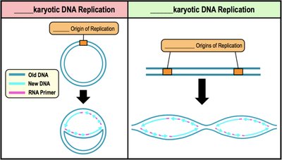
DNA replication begins at specific DNA sequences called origins of replication (ORI). Prokaryotes typically have a single, circular chromosome with one ORI, while eukaryotes have multiple, linear chromosomes with many ORIs.
Prokaryotic Replication: One origin, circular DNA.
Eukaryotic Replication: Multiple origins, linear DNA.
Replication Forks
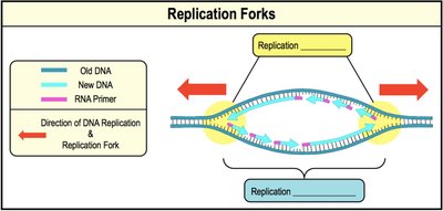
Proteins bind to the ORI and separate the two DNA strands, forming a replication bubble. Replication forks are Y-shaped regions at each end of the bubble where DNA is unwound and replication proceeds bidirectionally.
Replication Fork: Site where DNA is actively unwound and replicated.
Bidirectional Replication: DNA synthesis occurs in both directions from the origin.
Unwinding the DNA: Topoisomerase, Helicase & SSBs
Several proteins participate in the unwinding of DNA during replication:
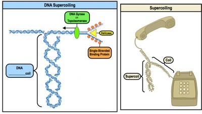
Topoisomerase: Cuts and rejoins DNA to relieve strain caused by supercoiling.
Helicase: Unwinds DNA by breaking hydrogen bonds, creating single-stranded DNA.
Single-Strand Binding Proteins (SSBs): Prevent reannealing and degradation of separated DNA strands.
Central Dogma of Molecular Biology
Overview of Central Dogma
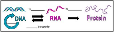
The central dogma describes the flow of genetic information from DNA to RNA to protein. Transcription is the process of synthesizing RNA from DNA, and translation is the process of synthesizing protein from RNA. Gene expression refers to the combined processes by which genotype becomes phenotype.
Transcription: DNA → RNA
Translation: RNA → Protein
Gene Expression: The full process by which genotype is expressed as phenotype.
Transcription
Introduction to Transcription
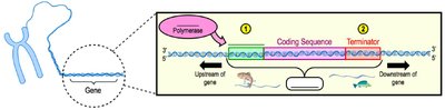
Transcription is the process of synthesizing RNA using DNA as a template. Genes are units of DNA that encode products such as proteins. Specific DNA sequences mark where transcription begins (promoter) and ends (terminator).
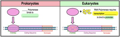
Promoter: DNA sequence where transcription starts; site of RNA polymerase attachment.
RNA Polymerase: Enzyme that synthesizes RNA from DNA template.
Terminator: DNA sequence where transcription ends.
Transcription Directionality
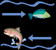
"Upstream" refers to DNA sequences before the start of transcription, while "downstream" refers to sequences after the start site. RNA polymerase moves along the template strand, synthesizing RNA in the 5' to 3' direction.
Overview of Transcription
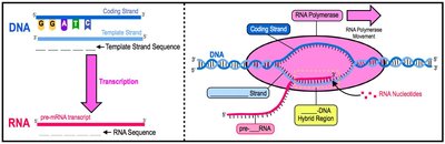
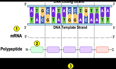
The two DNA strands in a gene are called the coding strand and the template strand. The RNA molecule has the same sequence as the coding strand (except T is replaced by U). Transcription builds RNA from the 5' to 3' end by pairing free RNA nucleotides with the DNA template.
Coding Strand: Has the same sequence as the RNA (except T/U).
Template Strand: Used by RNA polymerase to build RNA.
Base Pairing: A–U, C–G in RNA.
Steps of Transcription
Transcription consists of three main steps:
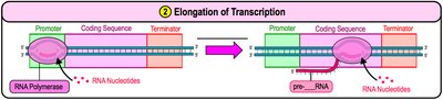
Initiation: RNA polymerase binds to the promoter and unwinds the DNA.
Elongation: RNA polymerase synthesizes RNA by pairing nucleotides with the DNA template.
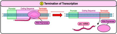
Termination: RNA polymerase reaches the terminator sequence and releases the RNA molecule.
Eukaryotic RNA Processing & Splicing
RNA Processing
Unlike prokaryotic mRNA, eukaryotic mRNA requires further modification after transcription. Pre-mRNA is processed by adding a 5' cap (modified guanine) and a poly-A tail (sequence of adenine nucleotides) to protect the mRNA and facilitate its export and translation.
5' Cap: Added to the 5' end; aids in stability and ribosome binding.
Poly-A Tail: Added to the 3' end; protects from degradation.


RNA Splicing
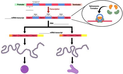
Eukaryotic genes contain introns (noncoding regions) and exons (coding regions). RNA splicing removes introns and joins exons to create mature mRNA. The spliceosome is responsible for this process. Alternative splicing allows a single gene to produce multiple products.
Introns: Noncoding regions, removed during splicing.
Exons: Coding regions, expressed in the final mRNA.
Spliceosome: Complex that removes introns.
Alternative Splicing: Generates diverse proteins from one gene.
Types of RNA
Major Types of RNA
Cells use several types of RNA, each with distinct functions:
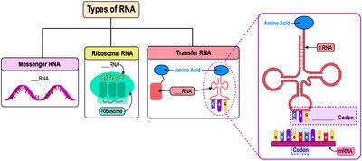
Messenger RNA (mRNA): Carries genetic information from DNA to ribosomes; contains codons.
Ribosomal RNA (rRNA): Forms part of ribosome structure.
Transfer RNA (tRNA): Brings amino acids to ribosome; contains anticodons complementary to mRNA codons.
Genetic Code
Understanding the Genetic Code
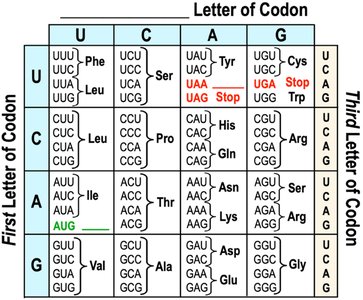
The genetic code is a set of rules by which DNA and RNA sequences are translated into proteins. It is nearly universal and reads three-nucleotide codons, each specifying an amino acid. The process involves converting DNA to mRNA, identifying codons, and translating them into amino acids until a stop codon is reached.
Codon: Three-nucleotide sequence in mRNA that codes for an amino acid.
Redundancy: Multiple codons can code for the same amino acid.
Start Codon: AUG (Methionine) initiates translation.
Stop Codons: UAA, UAG, UGA signal termination.
Translation
Introduction to Translation
Translation is the process of synthesizing proteins using the encoded messages of mRNA. Ribosomes facilitate translation, and tRNA brings amino acids to the ribosome, pairing its anticodon with the mRNA codon. Charged tRNA is attached to an amino acid, while discharged tRNA is not.
Ribosome: Site of protein synthesis.
tRNA: Transfers amino acids; contains anticodons.
Charged tRNA: tRNA with an attached amino acid.
*Additional info: The shape of tRNA is maintained by hydrogen bonding between base pairs of nucleotides.*
Ribosome Subunits and tRNA Binding Sites
Ribosomes consist of small and large subunits made of proteins and rRNA. Prokaryotes have 70S ribosomes (50S + 30S), while eukaryotes have 80S ribosomes (60S + 40S). Each ribosome has three tRNA binding sites:
A-site (Aminoacyl-tRNA): Holds tRNA with the next amino acid.
P-site (Peptidyl-tRNA): Holds tRNA with the growing polypeptide chain.
E-site (Exit): Discharged tRNAs leave the ribosome.
*Additional info: Ribosomes read mRNA in the 5' to 3' direction, and several ribosomes can translate the same mRNA simultaneously.*
Steps of Translation
Translation consists of three main steps:
Initiation: Small ribosomal subunit binds mRNA and tRNA; large subunit joins. The start codon (AUG) specifies Methionine.
Elongation: Amino acids are added one by one to the growing chain; ribosome moves along mRNA, tRNAs shift through A, P, and E sites.
Termination: Stop codon reaches A-site; release factors bind, polypeptide is released, and ribosome disassembles.
*Additional info: Peptide bonds are formed during elongation, just after a tRNA charged with the next amino acid binds to the A site.*
Post-Translational Modification
Types of Post-Translational Modifications (PTM)
After translation, proteins may undergo covalent modifications that regulate their activity. Common PTMs include methylation, acetylation, ubiquitination, phosphorylation, and glycosylation (addition of carbohydrates).
Glycosylation: Addition of carbohydrates to proteins.
Phosphorylation: Addition of phosphate groups; reversible and can activate/deactivate proteins.
Acetylation: Addition of acetyl groups.
Ubiquitination: Addition of ubiquitin, marking proteins for degradation.
Transcription vs. Translation: Review
Comparison and Contrast
Transcription and translation are distinct but interconnected processes in gene expression. Transcription converts DNA to RNA, while translation converts RNA to protein. The central dogma refers to the unidirectional flow of information: DNA → RNA → Protein.
*Additional info: DNA coding strand, template strand, and mRNA sequence can be determined using base pairing rules. The genetic code is used to translate mRNA into protein sequences.*
Mutations
Types and Effects of Mutations
Mutations are permanent changes in the DNA sequence. They can impact RNA and protein products, and may be harmful, beneficial, or neutral. Mutations can occur spontaneously or be induced by environmental factors (mutagens). They contribute to genetic diversity.
Frameshift Mutation: Insertion or deletion of nucleotides that alters the reading frame.
Missense Mutation: Substitution that changes one amino acid.
Nonsense Mutation: Substitution that creates a stop codon, terminating translation prematurely.
Silent Mutation: Substitution that does not change the amino acid.
*Additional info: Mutations in coding regions can affect protein function, while mutations in noncoding regions may have less impact.*
