 Indietro
Study Guide: Photosynthesis, Cell Division (Mitosis & Meiosis), and Mendelian Genetics
Guida di studio - Note intelligenti
 Appunti personalizzati basati sui tuoi materiali, ampliati con definizioni chiave, esempi e contesto.
Photosynthesis
Overview of Photosynthesis
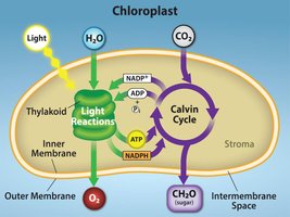
Photosynthesis is the process by which photoautotrophs (plants, algae, and some prokaryotes) convert solar energy into chemical energy stored in sugars and other organic molecules. This process occurs in chloroplasts and consists of two main phases: light-dependent reactions and light-independent reactions (Calvin cycle).
Light-dependent reactions: Occur in the thylakoid membrane, use light energy to produce ATP and NADPH.
Light-independent reactions (Calvin cycle): Occur in the stroma, use ATP and NADPH to fix CO2 into G3P (glyceraldehyde-3-phosphate).
Pigments: Chlorophyll a & b (green), carotenoids (yellow, orange, red), and vacuolar pigments (blue, violet, purple, dark red).
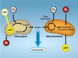
Connection Between Photosynthesis & Cellular Respiration
Photosynthesis and cellular respiration are interconnected processes. Photosynthesis stores energy in sugars, while cellular respiration releases that energy for cellular use.
Photosynthesis: Converts light energy to chemical energy, produces O2, and stores energy in sugars.
Cellular Respiration: Converts chemical energy in sugars to ATP, consumes O2, and releases CO2.
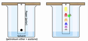
Chromatography in Photosynthesis
Chromatography is used to separate plant pigments based on their solubility and polarity. More oxygenated pigments are more polar.
Solvent: Petroleum ether + acetone.
Zeaxanthin: A xanthophyll pigment.
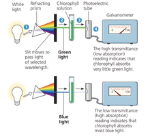
Absorption Spectra of Pigments
Pigments absorb specific wavelengths of light, which is measured using a spectrophotometer. The absorption spectra help determine which wavelengths are used in photosynthesis.
Absorbance: Indicates how much light is absorbed by a pigment at each wavelength.
Application: Pigments work together to utilize as much of the light spectrum as possible.
Cell Division
Somatic Cells vs. Gametes
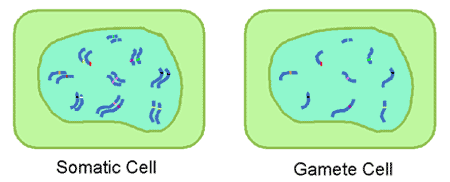
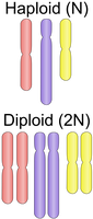
Human somatic cells are diploid (2n, 46 chromosomes), while gametes (sperm/ova) are haploid (n, 23 chromosomes). Fertilization fuses two haploid gametes to form a diploid zygote.
Somatic cells: Produced by mitosis.
Gametes: Produced by meiosis.
The Cell Cycle
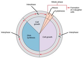
The cell cycle consists of interphase (G1, S, G2) and the mitotic phase (mitosis and cytokinesis).
G1: Cell growth.
S: DNA replication.
G2: Preparation for mitosis.
Mitosis: Chromosome separation.
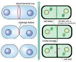
Cytokinesis: Cytoplasm division.
Cell Cycle Checkpoints
G1 checkpoint: Checks cell size, nutrients, DNA damage, and growth factors.
G2 checkpoint: Ensures complete DNA replication.
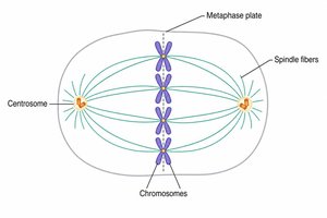
M checkpoint: Ensures chromosomes are properly aligned at the metaphase plate.
Mitosis
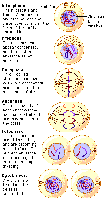
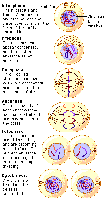
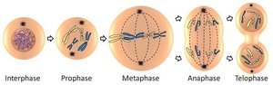
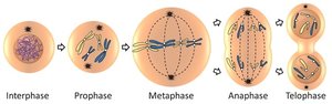
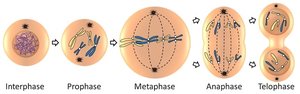
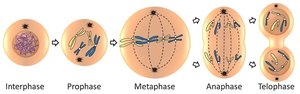
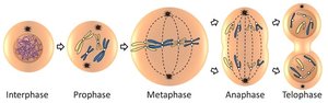
Mitosis produces two genetically identical daughter cells. The steps are:
Prophase: Chromatin condenses, nuclear membrane disappears, spindle forms.
Metaphase: Chromosomes align at the metaphase plate.
Anaphase: Sister chromatids separate and move to opposite poles.
Telophase: Nuclear envelope reforms, chromosomes uncoil.
Cytokinesis: Cytoplasm divides.
Chromosome Vocabulary
Chromosome: DNA and proteins during mitosis.
Chromatin: DNA and proteins during interphase.
Genome: All genetic information in a cell.
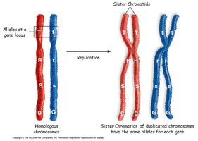
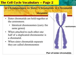
Sister chromatids: Duplicated chromosomes joined at the centromere.
Centromere: Region where kinetochore attaches.
Kinetochore: Protein structure for microtubule attachment.
Centrosome: Microtubule organizing center.
Centriole: Cylinder of microtubule triplets.
Meiosis
Purpose and Overview
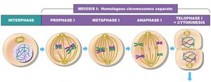
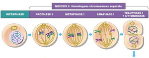
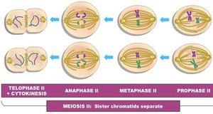
Meiosis reduces chromosome number by half and increases genetic diversity. It consists of two divisions:
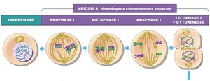
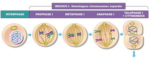
Meiosis I: Separates homologous chromosomes.
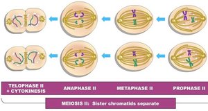
Meiosis II: Separates sister chromatids.
Crossing Over and Genetic Diversity
Crossing over occurs during prophase I of meiosis, where homologous chromosomes exchange genetic material at chiasmata, increasing genetic diversity. 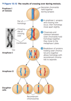
Meiosis I and II
Meiosis I: Homologous chromosomes align and are separated, resulting in haploid cells with sister chromatids.
Meiosis II: Sister chromatids are separated, resulting in four haploid cells.
Mendelian Genetics
Mendel’s Laws of Inheritance
Law of Segregation: Two alleles for a heritable character segregate during gamete formation, ending up in different gametes.
Law of Independent Assortment: Genes on different chromosomes assort independently during gamete formation.
Genetic Terminology
Locus: Location of a gene on a chromosome.
Allele: Different versions of a gene.
Homozygous: Two identical alleles (AA, BB).
Heterozygous: Two different alleles (Aa, Bb).
Punnett Square: Diagram used to predict offspring genotypes and phenotypes.
Dihybrid Crosses
Dihybrid crosses involve two genes, each with two alleles. The phenotypic ratio for a cross between two heterozygotes (RrYy x RrYy) is 9:3:3:1.
Round yellow: 9
Round green: 3
Wrinkled yellow: 3
Wrinkled green: 1
Step-by-Step Dihybrid Cross
Determine parental genotypes and possible gametes.
Combine parental genotypes to get F1 generation genotype.
Determine possible gametes from F1 generation.
Draw a 4x4 Punnett square using possible gametes.
Add possible F2 genotypes to the Punnett square.
Determine phenotypes from genotypes.
Additional info:
All equations and formulas are provided in LaTeX format as required.
Some context was inferred for completeness and clarity.
